Fraud Blocker Skip to main content

Home > All Images > 2008 > May > 23 May 2008

Images Dated 23rd May 2008 (page 6)

389 items

Background imageImages Dated 23rd May 2008: Europe, Germany

Europe, Germany
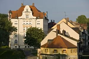
Germany, Bayern-Bavaria, Regensburg. Buildings on Wohrdstrasse by Danube River

Background imageImages Dated 23rd May 2008: Africa, Mauritius

Africa, Mauritius
Bamboo Grove, Sir Seewoosagur Ramgoolam Botanical Gardens, SSR Botanical Gardens or Royal Botanical Gardens near Pamplemousses, Northern Mauritius, Africa

Background imageImages Dated 23rd May 2008: Africa, Mauritius

Africa, Mauritius
Pond with giant Victoria amazonica water lillies, Sir Seewoosagur Ramgoolam Boatanical Gardens, Royal Botanical Gardens near Pamplemousses, Northern Mauritius, Africa

Background imageImages Dated 23rd May 2008: Africa, Mauritius

Africa, Mauritius
Pond with giant Victoria amazonica water lillies, Sir Seewoosagur Ramgoolam Boatanical Gardens, Royal Botanical Gardens near Pamplemousses, Northern Mauritius, Africa

Background imageImages Dated 23rd May 2008: Pond with giant Victoria amazonica water lillies, Sir Seewoosagur Ramgoolam Boatanical Gardens

Pond with giant Victoria amazonica water lillies, Sir Seewoosagur Ramgoolam Boatanical Gardens, Royal Botanical Gardens near Pamplemousses, Northern Mauritius, Africa

Background imageImages Dated 23rd May 2008: famous Catholic Church with Red Roof Notre Dame Auxiliaiatrice, cap Malheureux, Mauritius

famous Catholic Church with Red Roof Notre Dame Auxiliaiatrice, cap Malheureux, Mauritius, Africa

Background imageImages Dated 23rd May 2008: famous Catholic Church with Red Roof Notre Dame Auxiliatrice, cap Malheureux, Mauritius

famous Catholic Church with Red Roof Notre Dame Auxiliatrice, cap Malheureux, Mauritius, Africa

Background imageImages Dated 23rd May 2008: Grand Baie, Northern Mauritius, Africa

Grand Baie, Northern Mauritius, Africa

Background imageImages Dated 23rd May 2008: Grand Baie, Northern Mauritius, Africa

Grand Baie, Northern Mauritius, Africa

Background imageImages Dated 23rd May 2008: Sugar Cane, Lion Mountain in background, East Mauritius, Africa

Sugar Cane, Lion Mountain in background, East Mauritius, Africa

Background imageImages Dated 23rd May 2008: SCOTLAND: DUNKELD BRIDGE. View of Dunkeld Bridge, on the river Tay in Scotland

SCOTLAND: DUNKELD BRIDGE. View of Dunkeld Bridge, on the river Tay in Scotland. Wood engraving, c1875

Background imageImages Dated 23rd May 2008: Phagocytosis of a yeast spore, SEM

Phagocytosis of a yeast spore, SEM
Phagocytosis of a yeast spore. Coloured scanning electron micrograph (SEM) of a neutrophil (brown) phagocytosing (engulfing and destroying) a spore from the yeast Candida albicans (orange)

Background imageImages Dated 23rd May 2008: Phagocytosis of fungus spores, SEM

Phagocytosis of fungus spores, SEM
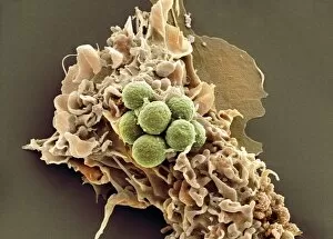
Phagocytosis of fungus spores. Coloured scanning electron micrograph (SEM) of a dendritic cell (brown) phagocytosing (engulfing and destroying) spores from the fungus Aspergillus fumigatus (green)

Background imageImages Dated 23rd May 2008: Phagocytosis of fungus spores, SEM

Phagocytosis of fungus spores, SEM
Phagocytosis of fungus spores. Coloured scanning electron micrograph (SEM) of a dendritic cell (blue) phagocytosing (engulfing and destroying) spores from the fungus Aspergillus fumigatus (red)

Background imageImages Dated 23rd May 2008: Phagocytosis of fungal spores, SEM

Phagocytosis of fungal spores, SEM
Phagocytosis of a fungus spore. Coloured scanning electron micrograph (SEM) of a neutrophil (brown) phagocytosing (engulfing and destroying) spores from the fungus Aspergillus fumigatus (red)

Background imageImages Dated 23rd May 2008: Phagocytosis of a fungus spore, SEM

Phagocytosis of a fungus spore, SEM
Phagocytosis of a fungus spore. Coloured scanning electron micrograph (SEM) of a spore from the fungus Aspergillus fumigatus (red) being phagocytosed (engulfed and destroyed)

Background imageImages Dated 23rd May 2008: Phagocytosis of fungal spores, SEM

Phagocytosis of fungal spores, SEM
Phagocytosis of a fungus spore. Coloured scanning electron micrograph (SEM) of a neutrophil (green) phagocytosing (engulfing and destroying) spores from the fungus Aspergillus fumigatus (yellow)

Background imageImages Dated 23rd May 2008: Phagocytosis of a fungus spore, SEM

Phagocytosis of a fungus spore, SEM
Phagocytosis of a fungus spore. Coloured scanning electron micrograph (SEM) of a spore from the fungus Aspergillus fumigatus (yellow) being phagocytosed (engulfed and destroyed)

Background imageImages Dated 23rd May 2008: Phagocytosis of fungus spores, SEM

Phagocytosis of fungus spores, SEM
Phagocytosis of fungus spores. Coloured scanning electron micrograph (SEM) of a dendritic cell (brown) phagocytosing (engulfing and destroying) spores from the fungus Aspergillus fumigatus (orange)

Background imageImages Dated 23rd May 2008: Phagocytosis of fungus spores, SEM

Phagocytosis of fungus spores, SEM
Phagocytosis of fungus spores. Coloured scanning electron micrograph (SEM) of a dendritic cell (cream) phagocytosing (engulfing and destroying) spores from the fungus Aspergillus fumigatus (red)

Background imageImages Dated 23rd May 2008: Influenza viruses, TEM

Influenza viruses, TEM
Influenza viruses. Coloured transmission electron micrograph (TEM) of influenza (flu) viruses (yellow) budding from a host cell. The host cell nucleus is black

Background imageImages Dated 23rd May 2008: White blood cells, SEM

White blood cells, SEM
White blood cells. Coloured scanning electron micrograph (SEM) of B lymphocyte white blood cells. Magnification: x5000 when printed at 10 centimetres wide

Background imageImages Dated 23rd May 2008: Water lily stem, SEM

Water lily stem, SEM
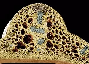
Water lily stem. Coloured scanning electron micrograph (SEM) of a freeze-fractured water lily stem showing numerous vascular bundles (grey) and large intercellular air spaces (holes)

Background imageImages Dated 23rd May 2008: Marjoram leaf surface, SEM

Marjoram leaf surface, SEM
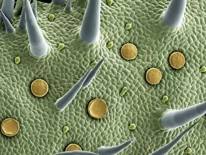
Marjoram (Origanum vulgare) leaf surface, coloured scanning electron micrograph (SEM). Glandular trichomes are yellow, hair-like trichomes are blue

Background imageImages Dated 23rd May 2008: White blood cells, SEM

White blood cells, SEM
White blood cells. Coloured scanning electron micrograph (SEM) of B lymphocyte white blood cells. Magnification: x5000 when printed at 10 centimetres wide

Background imageImages Dated 23rd May 2008: Skin, SEM

Skin, SEM
Skin. Coloured scanning electron micrograph (SEM) of squamous epithelial cells on the skin surface. Magnification: x350 when printed at 10 centimetres wide

Background imageImages Dated 23rd May 2008: Sperm cell, SEM

Sperm cell, SEM
Sperm cell. Coloured scanning electron micrograph (SEM) of a sperm cell in a fallopian tube. Magnification: x400 when printed at 10 centimetres wide

Background imageImages Dated 23rd May 2008: Human hair, SEM

Human hair, SEM
Human hair, coloured scanning electron micrograph (SEM). Magnification: x550 when printed at 10 centimetres wide

Background imageImages Dated 23rd May 2008: Skeletal muscle fibres, SEM

Skeletal muscle fibres, SEM
Skeletal muscle fibres, coloured scanning electron micrograph (SEM). Endomysial connective tissue is green. Magnification: x300 when printed at 10 centimetres wide

Background imageImages Dated 23rd May 2008: Baby spider, SEM

Baby spider, SEM
Baby spider, or spiderling, coloured scanning electron micrograph (SEM). Magnification: x25 when printed at 10 centimetres wide

Background imageImages Dated 23rd May 2008: Fat tissue, SEM

Fat tissue, SEM
Fat (adipose) tissue, coloured scanning electron micrograph (SEM). The fat cells (adipocytes, round) are surrounded by collagen fibres. Magnification: x650 when printed at 10 centimetres wide

Background imageImages Dated 23rd May 2008: Human hair, SEM

Human hair, SEM
Human hair, coloured scanning electron micrograph (SEM). Magnification: x550 when printed at 10 centimetres wide

Background imageImages Dated 23rd May 2008: Skin, SEM

Skin, SEM
Skin. Coloured scanning electron micrograph (SEM) of squamous epithelial cells on the skin surface. Magnification: x350 when printed at 10 centimetres wide

Background imageImages Dated 23rd May 2008: Gall bladder surface, SEM

Gall bladder surface, SEM
Internal lining of the gall bladder, coloured scanning electron micrograph (SEM). These columnar epithelial cells are covered in microvilli. Magnification: x2100 when printed at 10 centimetres wide

Background imageImages Dated 23rd May 2008: Trachea lining, SEM

Trachea lining, SEM
Trachea (wind pipe) lining, coloured scanning electron micrograph (SEM). Cilia are green and goblet cells (with microvilli) are purple. Magnification: x5000 when printed at 10 centimetres wide

Background imageImages Dated 23rd May 2008: Crenated red blood cells, SEM

Crenated red blood cells, SEM
Crenated red blood cells, coloured scanning electron micrograph (SEM). Magnification: x4000 when printed at 10 centimetres wide

Background imageImages Dated 23rd May 2008: Activated platelets, SEM

Activated platelets, SEM
Activated platelets, coloured scanning electron micrograph (SEM). Magnification: x6000 when printed at 10 centimetres wide

Background imageImages Dated 23rd May 2008: Kidney glomerulus, SEM

Kidney glomerulus, SEM
Kidney glomerulus, coloured scanning electron micrograph (SEM). Podocyte cells, which make up the surface of the glomerulus, are pink. Magnification: x650 when printed at 10 centimetres wide

Background imageImages Dated 23rd May 2008: Deformed sperm cell, SEM

Deformed sperm cell, SEM
Deformed sperm cell, coloured scanning electron micrograph (SEM). This cell has residual cytoplasm (lump, pink) below its head (red, top right)

Background imageImages Dated 23rd May 2008: Baby spider, SEM

Baby spider, SEM
Baby spider, or spiderling, coloured scanning electron micrograph (SEM). Magnification: x25 when printed at 10 centimetres wide

Background imageImages Dated 23rd May 2008: Fat tissue, SEM

Fat tissue, SEM
Fat (adipose) tissue, coloured scanning electron micrograph (SEM). The fat cells (adipocytes, round) are surrounded by collagen fibres. Lipid is green. Empty cells are seen at top right

Background imageImages Dated 23rd May 2008: Fat tissue, SEM

Fat tissue, SEM
Fat (adipose) tissue, coloured scanning electron micrograph (SEM). The fat cells (adipocytes, round) are surrounded by collagen fibres. Lipid is purple. Empty cells are seen at top right

Background imageImages Dated 23rd May 2008: Gall bladder surface, SEM

Gall bladder surface, SEM
Internal lining of the gall bladder, coloured scanning electron micrograph (SEM). These columnar epithelial cells are covered in microvilli. Magnification: x2100 when printed at 10 centimetres wide

Background imageImages Dated 23rd May 2008: Wood, SEM

Wood, SEM
Wood. Coloured scanning electron micrograph (SEM) of a section through wood showing the phloem vessels (dark green holes) and xylem tissue (lower frame)


All Professionally Made to Order for Quick Shipping


-

Why Choose Us?

We are a leading provider of Art Prints and Photo Gifts since 2003, working in partnership with a range of Sporting Clubs, Charities, Museums and Picture Libraries. A large share of profits from any of their images will go directly towards supporting that charity or club. Our archive of images is carefully curated to bring you a wide range of subjects, including landscapes, wildlife, architecture, and more. We ship from our partner labs in the UK, USA, EU (Netherlands) and Australia.
+

How do I place an order?

Ordering is quick & easy - Just follow 5 Simple Steps:
  1. Find your image: Use our search box or browse our online photo Collections to find the image you want.

  2. Choose your print format: Select your desired product and add it to your cart.

  3. Enter your details: If you're a returning customer, simply enter your email address and password, and we'll fill in your billing and shipping address details. All personal details are held securely and are fully GDPR compliant. As standard, we remove all Personally Identifiable Information after 12 months.

  4. Pay for your purchase: We use state-of-the-art security for online shopping and do not have access to your card details.

  5. Sit back and relax: We'll email you confirmation of your order and when it's dispatched. Registered customers can also track orders in the 'My Account' area.

+

How do I pay for an order?

You can pay for your order with most of the major credit and debit cards, or PayPal. For added security, major financial institutions process payment details separately and securely on our behalf. We do not have access to your online payment card details. Online payments are preferred however we do take phone orders. For UK customers only we accept cheques issued against a UK bank.
+

Is my data safe?

We take data security very seriously. We do not have access to your full card details and all payments are requested over a fully secure connection. Additionally, we fully comply with current European and GDPR legislation, and automatically remove all personal data after at most 12 months (unless you have an account with us and have signed in recently). We also have a strict opt-in policy and would never sell your personal details. Your data is only used to fulfill your order promptly and efficiently.
+

Is wall art ready to hang?

For quick and easy installation all wall art, including framed prints, canvas prints and metal prints are supplied with a ready to hang solution on the back. Generally, saw tooth hangers are applied as they allow wall art to hang flush against the wall. The serrated edge of the hanger prevents the frame from shifting or tilting when hung.
+

Are the photo prints fade resistant?

Yes, we use archival quality photo paper photographic paper for vivid reproduction Prints are an accurate representations of the original artwork, which is preserved for artistic character and authenticity. We guarantee they match previews shown on our web site