Fraud Blocker Skip to main content

Home > All Images > 2008 > November > 17 Nov 2008

Images Dated 17th November 2008 (page 5)

368 items

Background imageImages Dated 17th November 2008: Microhylid Frog - Tangkoko Nature Reserve - North Sulawesi - Indonesia

Microhylid Frog - Tangkoko Nature Reserve - North Sulawesi - Indonesia
MAR-1536 Microhylid Frog Tangkoko Nature Reserve - North Sulawesi - Indonesia Kaloula sp. Thomas Marent

Background imageImages Dated 17th November 2008: USA, Colorado, Pike National Forest. Bushy-tailed woodrat or packrat clings to the

USA, Colorado, Pike National Forest. Bushy-tailed woodrat or packrat clings to the side of a boulder in early morning light

Background imageImages Dated 17th November 2008: Thailand dessert tapioca flour jellies

Thailand dessert tapioca flour jellies
BMD-222 Thailand - dessert tapioca flour jellies Bangkok street food Brian McDairmant

Background imageImages Dated 17th November 2008: Central America, Costa Rica

Central America, Costa Rica
Costa Rica, Savegre River Valley. Juvenile magnificent hummingbird in rainstorm

Background imageImages Dated 17th November 2008: Red Telephone box, Nelsons Dockyard, Antigua, West Indies, Caribbean, Central

Red Telephone box, Nelsons Dockyard, Antigua, West Indies, Caribbean, Central America

Background imageImages Dated 17th November 2008: China, Suzhou. Cuisine of Suzhou

China, Suzhou. Cuisine of Suzhou

Background imageImages Dated 17th November 2008: China, Suzhou. Cuisine of Suzhou

China, Suzhou. Cuisine of Suzhou

Background imageImages Dated 17th November 2008: Asia, China

Asia, China
China, Suzhou. Temple of Mystery (Xuanmiaoguan)

Background imageImages Dated 17th November 2008: China, Suzhou. Candles at the Temple of Mystery (Xuanmiaoguan)

China, Suzhou. Candles at the Temple of Mystery (Xuanmiaoguan)

Background imageImages Dated 17th November 2008: Asia, China

Asia, China
China, Suzhou. The Institute of Su Zhou Embroidery

Background imageImages Dated 17th November 2008: Asia, China

Asia, China
China, Suzhou. Barges on the Grand Canal

Background imageImages Dated 17th November 2008: Asia, China

Asia, China
China, Zhouzhuang. Prayer ribbons Quanfu Temple

Background imageImages Dated 17th November 2008: China, Zhouzhuang. Quanfu Temple

China, Zhouzhuang. Quanfu Temple

Background imageImages Dated 17th November 2008: Asia, China

Asia, China
China, Zhouzhuang. Quanfu Temple

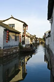
Background imageImages Dated 17th November 2008: China, Zhouzhuang. Historic Zhouzhuang Water Village

China, Zhouzhuang. Historic Zhouzhuang Water Village

Background imageImages Dated 17th November 2008: China, Zhouzhuang. Historic Zhouzhuang Water Village

China, Zhouzhuang. Historic Zhouzhuang Water Village

Background imageImages Dated 17th November 2008: Asia, China

Asia, China
China, Zhouzhuang. Historic Zhouzhuang Water Village

Background imageImages Dated 17th November 2008: Asia, China

Asia, China
China, Zhouzhuang. Historic Zhouzhuang Water Village

Background imageImages Dated 17th November 2008: China, Zhouzhuang. Historic Zhouzhuang Water Village

China, Zhouzhuang. Historic Zhouzhuang Water Village

Background imageImages Dated 17th November 2008: Africa, Kenya

Africa, Kenya
Group of Topi, Damaliscus korrigum, silhouetted at sunset in the Masai Mara Game Reserve, Kenya

Background imageImages Dated 17th November 2008: Africa, Kenya

Africa, Kenya
Cheetah, Acinonyx jubatus, with cub in the Masai Mara GR, Kenya

Background imageImages Dated 17th November 2008: Africa, Kenya

Africa, Kenya
Group of Topi, Damaliscus korrigum, silhouetted at sunset in the Masai Mara Game Reserve, Kenya

Background imageImages Dated 17th November 2008: Paramecium protozoan, SEM

Paramecium protozoan, SEM
Paramecium protozoan, coloured scanning electron micrograph (SEM). Paramecia are a group of unicellular ciliate protozoa. They inhabit fresh water, and feed mainly on bacteria and smaller protozoa

Background imageImages Dated 17th November 2008: Thyroid parafollicular cell, TEM

Thyroid parafollicular cell, TEM
Thyroid parafollicular cell, coloured transmission electron micrograph (TEM). This section has revealed the cells nucleus (light brown)

Background imageImages Dated 17th November 2008: Tooth enamel formation, SEM

Tooth enamel formation, SEM
Tooth enamel formation. Coloured scanning electron micrograph (SEM) of a freeze-fractured section through a tooth, showing the enamel-forming cell layer (blue)

Background imageImages Dated 17th November 2008: Tooth enamel formation, SEM

Tooth enamel formation, SEM
Tooth enamel formation. Coloured scanning electron micrograph (SEM) of a freeze-fractured section through a tooth, showing the enamel-forming cell layer (blue)

Background imageImages Dated 17th November 2008: Rosemary leaf structure, SEM

Rosemary leaf structure, SEM
Rosemary leaf. Coloured scanning electron micrograph (SEM) of a freeze-fracture through a leaf from a rosemary plant (Rosmarinus officinalis). The fracture has revealed vascular tissue (green)

Background imageImages Dated 17th November 2008: Plasma cell, TEM

Plasma cell, TEM
Plasma cell, coloured transmission electron micrograph (TEM). This section has revealed the cells large central nucleus (purple)

Background imageImages Dated 17th November 2008: Pancreas blood vessel, light micrograph

Pancreas blood vessel, light micrograph
Pancreas blood vessel. Light micrograph of a section through a blood vessel in the pancreas, showing red blood cells (red). Surrounding the blood vessel are pancreatic exocrine cells (brown)

Background imageImages Dated 17th November 2008: Asteroid near Mars, artwork

Asteroid near Mars, artwork
Asteroid near Mars, computer artwork. Mars is the fourth planet from the Sun. It is scarred by around 43, 000 impact craters that have a diameter of over 5 kilometres

Background imageImages Dated 17th November 2008: Oviduct mucosal folds, light micrograph

Oviduct mucosal folds, light micrograph. This section through the folds shows the ciliated columnar epithelium (red) and the connective tissue core (green)

Background imageImages Dated 17th November 2008: Athletes foot fungus, SEM

Athletes foot fungus, SEM
Athletes foot. Coloured scanning electron micrograph (SEM) of spores (pink) of the fungus that causes athletes foot (tinea pedis) on skin (brown) from a human foot

Background imageImages Dated 17th November 2008: Intestinal endocrine cell, TEM

Intestinal endocrine cell, TEM
Intestinal endocrine cell, coloured transmission electron micrograph (TEM). This is a type 1 intestinal endocrine cell. The granules (green) contain serotonin

Background imageImages Dated 17th November 2008: Ariane 5 rocket

Ariane 5 rocket. 8 meter high model of the Ariane 5 rocket, an expendable (single use) launch system. It was designed to place space probes

Background imageImages Dated 17th November 2008: Pancreas acinus, SEM

Pancreas acinus, SEM
Pancreas acinus. Coloured scanning electron micrograph (SEM) of a freeze-fracture through an acinus (yellow) in the pancreas. An acinus is a collection of glandular epithelial cells

Background imageImages Dated 17th November 2008: Giovanni Schiaparelli, Italian astronomer

Giovanni Schiaparelli, Italian astronomer
Giovanni Virginio Schiaparelli (1835-1910), Italian astronomer. Schiaparelli was, from 1862, the Director of the Milan Observatory for forty years

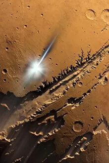
Background imageImages Dated 17th November 2008: Asteroid impact on Mars, artwork

Asteroid impact on Mars, artwork
Asteroid impact on Mars, computer artwork. Mars is the fourth planet from the Sun. It is scarred by around 43, 000 impact craters that have a diameter of over 5 kilometres

Background imageImages Dated 17th November 2008: Motor neurons, light micrograph

Motor neurons, light micrograph. Motor neurons are responsible for passing information around the central nervous system (CNS) and from the CNS to the rest of the body

Background imageImages Dated 17th November 2008: Yeast cells, SEM

Yeast cells, SEM
Yeast cells. Coloured scanning electron micrograph (SEM) of cells of bakers yeast (Saccharomyces cerevisiae) from part of a dried, commercial yeast pellet

Background imageImages Dated 17th November 2008: Paramecium protozoan, SEM

Paramecium protozoan, SEM
Paramecium protozoan, coloured scanning electron micrograph (SEM). Paramecia are a group of unicellular ciliate protozoa. They inhabit fresh water, and feed mainly on bacteria and smaller protozoa

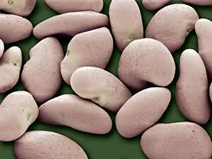
Background imageImages Dated 17th November 2008: Alfalfa seeds

Alfalfa seeds. Coloured scanning electron micrograph (SEM) of Alfafa (Medicago sativa) seeds. Alfalfa, also known as lucerne, is cultivated as an important forage crop (eaten by grazing livestock)

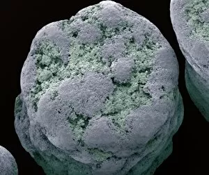
Background imageImages Dated 17th November 2008: Dried commercial yeast pellet, SEM

Dried commercial yeast pellet, SEM
Dried commercial yeast pellet. Coloured scanning electron micrograph (SEM) of a dried pellet of bakers yeast (Saccharomyces cerevisiae). Yeast is a fungus and consists of single vegetative cells. S

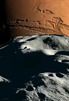
Background imageImages Dated 17th November 2008: Mars as seen from Phobos, artwork

Mars as seen from Phobos, artwork
Mars as seen from Phobos, computer artwork. Phobos (grey, foreground) is the larger of the two Martian moons. It is irregular in shape, measuring 19 by 21 by 27 kilometres

Background imageImages Dated 17th November 2008: Dried commercial yeast pellet, SEM

Dried commercial yeast pellet, SEM
Dried commercial yeast pellet. Coloured scanning electron micrograph (SEM) of a dried pellet of bakers yeast (Saccharomyces cerevisiae). Yeast is a fungus and consists of single vegetative cells. S

Background imageImages Dated 17th November 2008: Soviet Proton rocket

Soviet Proton rocket. This rocket (lower centre) is an expendable (single use) system for both commercial and Russian government launches

Background imageImages Dated 17th November 2008: Thyroid parafollicular cell, TEM

Thyroid parafollicular cell, TEM
Thyroid parafollicular cell, coloured transmission electron micrograph (TEM). This section has revealed the cells nucleus (green)

Background imageImages Dated 17th November 2008: Athletes foot fungus, SEM

Athletes foot fungus, SEM
Athletes foot. Coloured scanning electron micrograph (SEM) of spores (yellow) of the fungus that causes athletes foot (tinea pedis) on skin (purple) from a human foot

Background imageImages Dated 17th November 2008: Osteoclasts in bone lacunae, SEM

Osteoclasts in bone lacunae, SEM
Osteoclasts in bone lacunae, coloured scanning electron micrograph (SEM). These osteoclasts are seen in Howslips lacunae, spaces in the bone where they are responsible for remodeling the bone


All Professionally Made to Order for Quick Shipping


-

Why Choose Us?

We are a leading provider of Art Prints and Photo Gifts since 2003, working in partnership with a range of Sporting Clubs, Charities, Museums and Picture Libraries. A large share of profits from any of their images will go directly towards supporting that charity or club. Our archive of images is carefully curated to bring you a wide range of subjects, including landscapes, wildlife, architecture, and more. We ship from our partner labs in the UK, USA, EU (Netherlands) and Australia.
+

How do I place an order?

Ordering is quick & easy - Just follow 5 Simple Steps:
  1. Find your image: Use our search box or browse our online photo Collections to find the image you want.

  2. Choose your print format: Select your desired product and add it to your cart.

  3. Enter your details: If you're a returning customer, simply enter your email address and password, and we'll fill in your billing and shipping address details. All personal details are held securely and are fully GDPR compliant. As standard, we remove all Personally Identifiable Information after 12 months.

  4. Pay for your purchase: We use state-of-the-art security for online shopping and do not have access to your card details.

  5. Sit back and relax: We'll email you confirmation of your order and when it's dispatched. Registered customers can also track orders in the 'My Account' area.

+

How do I pay for an order?

You can pay for your order with most of the major credit and debit cards, or PayPal. For added security, major financial institutions process payment details separately and securely on our behalf. We do not have access to your online payment card details. Online payments are preferred however we do take phone orders. For UK customers only we accept cheques issued against a UK bank.
+

Is my data safe?

We take data security very seriously. We do not have access to your full card details and all payments are requested over a fully secure connection. Additionally, we fully comply with current European and GDPR legislation, and automatically remove all personal data after at most 12 months (unless you have an account with us and have signed in recently). We also have a strict opt-in policy and would never sell your personal details. Your data is only used to fulfill your order promptly and efficiently.
+

Is wall art ready to hang?

For quick and easy installation all wall art, including framed prints, canvas prints and metal prints are supplied with a ready to hang solution on the back. Generally, saw tooth hangers are applied as they allow wall art to hang flush against the wall. The serrated edge of the hanger prevents the frame from shifting or tilting when hung.
+

Are the photo prints fade resistant?

Yes, we use archival quality photo paper photographic paper for vivid reproduction Prints are an accurate representations of the original artwork, which is preserved for artistic character and authenticity. We guarantee they match previews shown on our web site