Fraud Blocker Skip to main content

Home > All Images > 2012 > January > 11 Jan 2012

Images Dated 11th January 2012 (page 6)

363 items

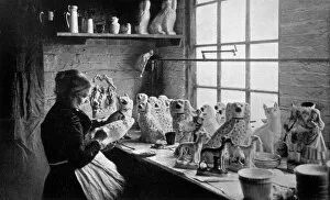
Background imageImages Dated 11th January 2012: Making Ceramic Dogs

Making Ceramic Dogs
A woman puts the finishing touches to ceramic dogs, popular cottage ornaments. Date: circa 1900

Background imageImages Dated 11th January 2012: Farmer in Autumn 1906

Farmer in Autumn 1906
An American Farmer in Autumn Date: 1906

Background imageImages Dated 11th January 2012: French Victory / Algeria

French Victory / Algeria
A French victory over the nationalists in southern Algeria au profit de nos interets commerciaux et de notre prestige moral... Date: Spring 1897

Background imageImages Dated 11th January 2012: Picking Fruit in America

Picking Fruit in America
Girls and boys gathering berries in America. Date: 1874

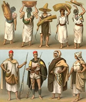
Background imageImages Dated 11th January 2012: Racial / N Africa / Kabyles

Racial / N Africa / Kabyles
Various Kabyles of Algeria and Tunisia, male and female Date: mid-19th century

Background imageImages Dated 11th January 2012: Ferdinand Duc Orleans

Ferdinand Duc Orleans
FERDINAND PHILLIPPE duc d ORLEANS son of Louis Philippe, military commander in Algeria. Date: 1810 - 1842

Background imageImages Dated 11th January 2012: Tobacco industry in Algiers

Tobacco industry in Algiers
Algeria : delivering tobacco to the shop of Hussein-Dey, Algiers Date: 1862

Background imageImages Dated 11th January 2012: Billy Wright - Wolverhampton Wanderers

Billy Wright - Wolverhampton Wanderers
Football - 1951 / 1952 season Wolverhampton Wanderers captain Billy Wright leads his team out

Background imageImages Dated 11th January 2012: Frog skin, SEM

Frog skin, SEM
Frog skin. Coloured scanning electron micrograph (SEM) of the skin of a frog, showing numerous folds (microplicae). These folds maximise the surface area to allow for increased cutaneous gas

Background imageImages Dated 11th January 2012: Intestinal papillomas, SEM

Intestinal papillomas, SEM
Intestinal papillomas. Coloured scanning electron micrograph (SEM) of a papilloma from a persons intestine. Papillomas are benign (non-cancerous) tumours that arise from epithelial (lining) tissue

Background imageImages Dated 11th January 2012: Frog pigment cell, SEM

Frog pigment cell, SEM
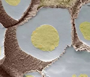
Frog pigment cell. Coloured scanning electron micrograph (SEM) of a section through a chromatophore pigment cell from the skin of a frog

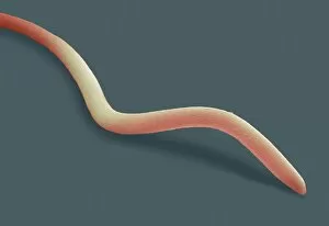
Background imageImages Dated 11th January 2012: Nematode worm, SEM

Nematode worm, SEM
Nematode worm. Coloured scanning electron micrograph (SEM) of a nematode. Nematodes, or roundworms, are one of the most diverse of all animals. Magnification: x270 when printed 10 centimetres wide

Background imageImages Dated 11th January 2012: Frog pigment cell, SEM

Frog pigment cell, SEM
Frog pigment cell. Coloured scanning electron micrograph (SEM) of a section through a chromatophore pigment cell from the skin of a frog

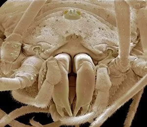
Background imageImages Dated 11th January 2012: Harvestman spider, SEM

Harvestman spider, SEM
Harvestman spider. Coloured scanning electron micrograph (SEM) of the head of a harvestman (order Opiliones), showing its large mandibles (jaws, lower centre)

Background imageImages Dated 11th January 2012: Antlion larva, SEM

Antlion larva, SEM
Antlion larva, coloured scanning electron micrograph (SEM). The larval form of this insect (family Myrmeleontide) lives at the bottom of a cone shaped hole

Background imageImages Dated 11th January 2012: Ant head, SEM

Ant head, SEM
Ant head. Coloured scanning electron micrograph (SEM) of the head of an ant (family Formicidae) found on the island of Cyprus, showing its large compound eyes (orange)

Background imageImages Dated 11th January 2012: Ant head, SEM

Ant head, SEM
Ant head. Coloured scanning electron micrograph (SEM) of the head of an ant (family Formicidae) found on the island of Cyprus, showing its large compound eyes (orange)

Background imageImages Dated 11th January 2012: Mosquito head, SEM

Mosquito head, SEM
Mosquito head. Coloured scanning electron micrograph (SEM) of the head of a mosquito, showing its large compound eyes. Each eye consists of many lenses (spheres) known as ommatidia

Background imageImages Dated 11th January 2012: Harvestman spider, SEM

Harvestman spider, SEM
Harvestman spider. Coloured scanning electron micrograph (SEM) of the head of a harvestman (order Opiliones), showing its large mandibles (jaws, lower centre)

Background imageImages Dated 11th January 2012: Vorticella protozoan, SEM

Vorticella protozoan, SEM
Vorticella protozoan. Coloured scanning electron micrograph (SEM) of a Vorticella sp. ciliate protozoan. This single-celled organism consists of a bell-shaped head

Background imageImages Dated 11th January 2012: Moth head, SEM

Moth head, SEM
Moth head. Coloured scanning electron micrograph (SEM) of the head of a moth, showing one of its two large compound eyes (purple) and its long proboscis (coiled)

Background imageImages Dated 11th January 2012: Mosquito head, SEM

Mosquito head, SEM
Mosquito head. Coloured scanning electron micrograph (SEM) of the head of a mosquito, showing its large compound eyes. Each eye consists of many lenses (spheres) known as ommatidia

Background imageImages Dated 11th January 2012: Frogspawn, SEM

Frogspawn, SEM
Frogspawn. Coloured scanning electron micrograph (SEM) of a section through frog eggs, showing the large nucleus containing nucleoli, surrounded by yolk

Background imageImages Dated 11th January 2012: Frog skin, SEM

Frog skin, SEM
Frog skin. Coloured scanning electron micrograph (SEM) of the skin of a frog, showing numerous folds (microplicae). These folds maximise the surface area to allow for increased cutaneous gas

Background imageImages Dated 11th January 2012: Nasal epithelium, SEM

Nasal epithelium, SEM
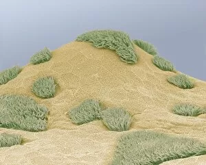
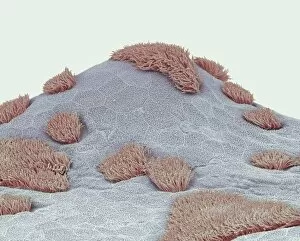
Nasal epithelium. Coloured scanning electron micrograph (SEM) of the lining of the nasal cavity. The nasal cavity contains both ciliated and non-ciliated (secretory) cells

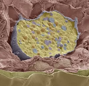
Background imageImages Dated 11th January 2012: Pancreas tissue, SEM

Pancreas tissue, SEM
Pancreas tissue. Coloured scanning electron micrograph (SEM) of fractured pancreas tissue, showing numerous acinar cells, containing secretory zymogen granules

Background imageImages Dated 11th January 2012: Antlion larva, SEM

Antlion larva, SEM
Antlion larva, coloured scanning electron micrograph (SEM). The larval form of this insect (family Myrmeleontide) lives at the bottom of a cone shaped hole

Background imageImages Dated 11th January 2012: Pancreas tissue, SEM

Pancreas tissue, SEM
Pancreas tissue. Coloured scanning electron micrograph (SEM) of fractured pancreas tissue, showing numerous acinar cells, containing secretory zymogen granules

Background imageImages Dated 11th January 2012: Intestinal papilloma, SEM

Intestinal papilloma, SEM
Intestinal papilloma. Coloured scanning electron micrograph (SEM) of a papilloma from a persons intestine. Papillomas are benign (non-cancerous) tumours that arise from epithelial (lining) tissue

Background imageImages Dated 11th January 2012: Nasal epithelium, SEM

Nasal epithelium, SEM
Nasal epithelium. Coloured scanning electron micrograph (SEM) of the lining of the nasal cavity. The nasal cavity contains both ciliated and non-ciliated (secretory) cells

Background imageImages Dated 11th January 2012: Osteoblast bone cell, SEM

Osteoblast bone cell, SEM
Osteoblast bone cell. Coloured scanning electron micrograph (SEM) of an osteoblast (centre) in bone tissue. Osteoblasts are primarily found in regions of new bone growth

Background imageImages Dated 11th January 2012: Intestinal papilloma, SEM

Intestinal papilloma, SEM
Intestinal papilloma. Coloured scanning electron micrograph (SEM) of a papilloma from a persons intestine. Papillomas are benign (non-cancerous) tumours that arise from epithelial (lining) tissue

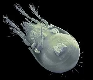
Background imageImages Dated 11th January 2012: Soil mite, SEM

Soil mite, SEM
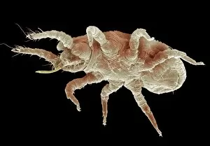
Soil mite. Coloured scanning electron micrograph (SEM) of a soil mite. This mite is typically found in moss and leaf litter

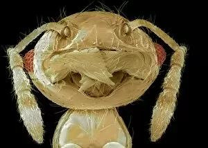
Background imageImages Dated 11th January 2012: Tropical ant head, SEM

Tropical ant head, SEM
Tropical ant head. Coloured scanning electron micrograph (SEM) of the head of a tropical ant (family Formicidae), showing one of its compound eyes (red)

Background imageImages Dated 11th January 2012: Ichthyostega, artwork

Ichthyostega, artwork
Ichthyostega. Computer artwork of an Ichthyostega around 350 million years ago during the Late Devonian period, in what is now the Canadian Arctic

Background imageImages Dated 11th January 2012: Thrip head, SEM

Thrip head, SEM
Thrip head. Coloured scanning electron micrograph (SEM) of the head of a thrip, showing its large compound eyes. Thrips (order Thysanoptera)

Background imageImages Dated 11th January 2012: Tropical ant head, SEM

Tropical ant head, SEM
Tropical ant head. Coloured scanning electron micrograph (SEM) of the head of a tropical ant (family Formicidae), showing its large mandibles (jaws, centre)

Background imageImages Dated 11th January 2012: Thrip, SEM

Thrip, SEM
Thrip, coloured scanning electron micrograph (SEM). Thrips (order Thysanoptera) feed on sap by piercing plant cells with their paired maxillary stylets (not seen), which form a feeding tube

Background imageImages Dated 11th January 2012: Ichthyostega, artwork

Ichthyostega, artwork
Ichthyostega. Computer artwork of a submerged Ichthyostega about to eat a drowned centipede in a floodplain, around 350 million years ago during the Late Devonian period

Background imageImages Dated 11th January 2012: Tropical ant head, SEM

Tropical ant head, SEM
Tropical ant head. Coloured scanning electron micrograph (SEM) of the head of a tropical ant (family Formicidae), showing one of its compound eyes (red)

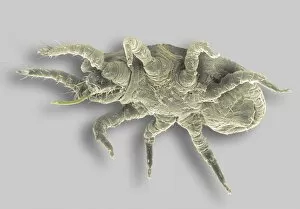
Background imageImages Dated 11th January 2012: Soil mite, SEM

Soil mite, SEM
Soil mite. Coloured scanning electron micrograph (SEM) of a soil mite. This mite is typically found in moss and leaf litter

Background imageImages Dated 11th January 2012: Next generation crew capsule, artwork

Next generation crew capsule, artwork
Next generation crew capsule. Computer artwork showing a next generation Deep Space Vehicles (DSV) crew capsule (centre right) mated with an Extended Stay Module (ESM)

Background imageImages Dated 11th January 2012: Vorticella protozoan, SEM

Vorticella protozoan, SEM
Vorticella protozoan. Coloured scanning electron micrograph (SEM) of a Vorticella sp. ciliate protozoan. This single-celled organism consists of a bell-shaped head

Background imageImages Dated 11th January 2012: Thrip, SEM

Thrip, SEM
Thrip, coloured scanning electron micrograph (SEM). Thrips (order Thysanoptera) feed on sap by piercing plant cells with their paired maxillary stylets (not seen), which form a feeding tube

Background imageImages Dated 11th January 2012: Soil mite, SEM

Soil mite, SEM
Soil mite. Coloured scanning electron micrograph (SEM) of a soil mite. This mite is typically found in moss and leaf litter

Background imageImages Dated 11th January 2012: Tropical ant head, SEM

Tropical ant head, SEM
Tropical ant head. Coloured scanning electron micrograph (SEM) of the head of a tropical ant (family Formicidae), showing its compound eyes (red) and large mandibles (jaws, centre)

Background imageImages Dated 11th January 2012: Thrip head, SEM

Thrip head, SEM
Thrip head. Coloured scanning electron micrograph (SEM) of the head of a thrip, showing its large compound eyes. Thrips (order Thysanoptera)

Background imageImages Dated 11th January 2012: Soil mite, SEM

Soil mite, SEM
Soil mite. Coloured scanning electron micrograph (SEM) of a soil mite. This mite is typically found in moss and leaf litter


All Professionally Made to Order for Quick Shipping


-

Why Choose Us?

We are a leading provider of Art Prints and Photo Gifts since 2003, working in partnership with a range of Sporting Clubs, Charities, Museums and Picture Libraries. A large share of profits from any of their images will go directly towards supporting that charity or club. Our archive of images is carefully curated to bring you a wide range of subjects, including landscapes, wildlife, architecture, and more. We ship from our partner labs in the UK, USA, EU (Netherlands) and Australia.
+

How do I place an order?

Ordering is quick & easy - Just follow 5 Simple Steps:
  1. Find your image: Use our search box or browse our online photo Collections to find the image you want.

  2. Choose your print format: Select your desired product and add it to your cart.

  3. Enter your details: If you're a returning customer, simply enter your email address and password, and we'll fill in your billing and shipping address details. All personal details are held securely and are fully GDPR compliant. As standard, we remove all Personally Identifiable Information after 12 months.

  4. Pay for your purchase: We use state-of-the-art security for online shopping and do not have access to your card details.

  5. Sit back and relax: We'll email you confirmation of your order and when it's dispatched. Registered customers can also track orders in the 'My Account' area.

+

How do I pay for an order?

You can pay for your order with most of the major credit and debit cards, or PayPal. For added security, major financial institutions process payment details separately and securely on our behalf. We do not have access to your online payment card details. Online payments are preferred however we do take phone orders. For UK customers only we accept cheques issued against a UK bank.
+

Is my data safe?

We take data security very seriously. We do not have access to your full card details and all payments are requested over a fully secure connection. Additionally, we fully comply with current European and GDPR legislation, and automatically remove all personal data after at most 12 months (unless you have an account with us and have signed in recently). We also have a strict opt-in policy and would never sell your personal details. Your data is only used to fulfill your order promptly and efficiently.
+

Is wall art ready to hang?

For quick and easy installation all wall art, including framed prints, canvas prints and metal prints are supplied with a ready to hang solution on the back. Generally, saw tooth hangers are applied as they allow wall art to hang flush against the wall. The serrated edge of the hanger prevents the frame from shifting or tilting when hung.
+

Are the photo prints fade resistant?

Yes, we use archival quality photo paper photographic paper for vivid reproduction Prints are an accurate representations of the original artwork, which is preserved for artistic character and authenticity. We guarantee they match previews shown on our web site