Fraud Blocker Skip to main content

Home > All Images > 2012 > November > 1 Nov 2012

Images Dated 1st November 2012 (page 5)

303 items

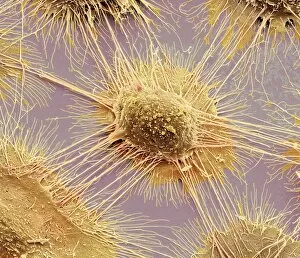
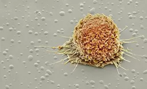
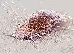
Background imageImages Dated 1st November 2012: Mouth cancer cell, SEM

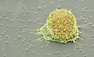
Mouth cancer cell, SEM
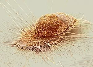
Mouth cancer cell. Coloured scanning electron micrograph (SEM) of a squamous cell carcinoma (cancer) cell from a human mouth

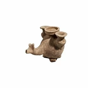
Background imageImages Dated 1st November 2012: Pre Columbian zoomorphic

Pre Columbian zoomorphic

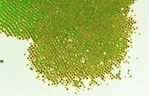
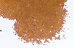
Background imageImages Dated 1st November 2012: Phosphate crystal, SEM

Phosphate crystal, SEM
Coloured scanning electron micrograph (SEM) of a single phosphate crystal. A phosphate is an inorganic chemical and a salt of phosphoric acid

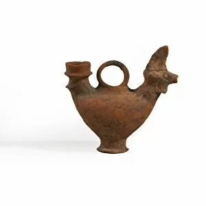
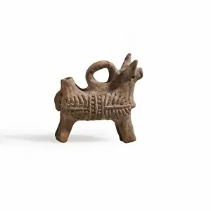
Background imageImages Dated 1st November 2012: Zoomorphic terracotta vessel

Zoomorphic terracotta vessel Byzantine period 5-7th century CE

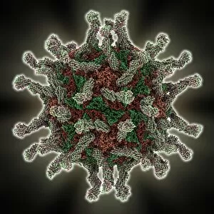
Background imageImages Dated 1st November 2012: Human poliovirus, molecular model

Human poliovirus, molecular model
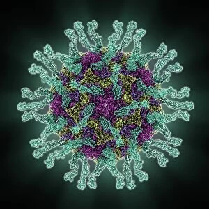
Human poliovirus particle. Computer model of the capsid of the human poliovirus. The capsid is a protein coat that encloses the viruss genetic information (genome), stored as RNA (ribonucleic acid)

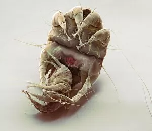
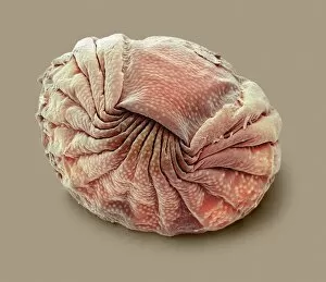
Background imageImages Dated 1st November 2012: Mange mites, SEM

Mange mites, SEM
Mange mites. Coloured scanning electron micrograph (SEM) of two mites of different sizes belonging to the family Sarcoptidae

Background imageImages Dated 1st November 2012: Mange mites, SEM

Mange mites, SEM
Mange mites. Coloured scanning electron micrograph (SEM) of two mites of different sizes belonging to the family Sarcoptidae

Background imageImages Dated 1st November 2012: Developing German wasp in a brood cell

Developing German wasp in a brood cell
A single brood cell within a nest of the German wasp (Vespula germanica). The nest is initiated in Spring by a fertilised female queen wasp

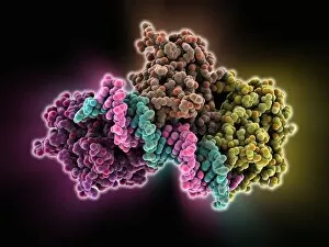
Background imageImages Dated 1st November 2012: Tumour suppressor protein molecular model

Tumour suppressor protein molecular model
Tumour suppressor protein. Molecular model of the tumour suppressor protein p53 (blue and purple) bound to a molecule of DNA (deoxyribonucleic acid, yellow and pink)

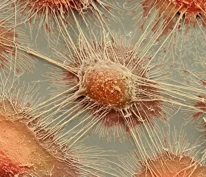
Background imageImages Dated 1st November 2012: Mouth cancer cells, SEM

Mouth cancer cells, SEM
Mouth cancer cells. Coloured scanning electron micrograph (SEM) of squamous cell carcinoma (cancer) cells from a human mouth

Background imageImages Dated 1st November 2012: Flu virus surface protein molecule

Flu virus surface protein molecule. Computer model showing the molecular structure of the neuraminidase glycoprotein enzyme found on the surface of the influenza A (flu) virus

Background imageImages Dated 1st November 2012: Intestinal brush border, TEM

Intestinal brush border, TEM
Intestinal brush border. Transmission electron micrograph (TEM) of a section through a brush border (centre) in the small intestine

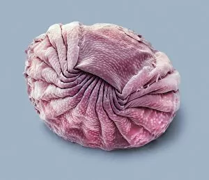
Background imageImages Dated 1st November 2012: Bdelloid rotifer, SEM

Bdelloid rotifer, SEM
Bdelloid rotifer. Coloured scanning electron micrograph (SEM) of a rotifer (class Bdelloidea) from a pond. Rotifers (phylum Rotifera) are microscopic aquatic animals that are related to roundworms

Background imageImages Dated 1st November 2012: Greek terracotta oil jug C014 / 6425

Greek terracotta oil jug C014 / 6425
Lekythos style Greek terracotta oil jug 4th century BCE

Background imageImages Dated 1st November 2012: Rhodopsin molecule

Rhodopsin molecule. Computer model showing the structure of a molecule of the rhodopsin complex. Rhodopsin (also called visual purple)

Background imageImages Dated 1st November 2012: Mouth cancer cell, SEM

Mouth cancer cell, SEM
Mouth cancer cell. Coloured scanning electron micrograph (SEM) of a squamous cell carcinoma (cancer) cell from a human mouth

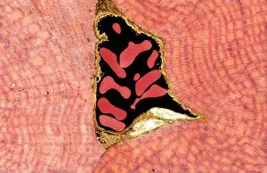
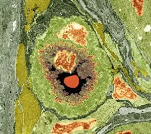
Background imageImages Dated 1st November 2012: Muscle arteriole, TEM

Muscle arteriole, TEM
Muscle arteriole. Transmission electron micrograph (TEM) of a section through an arteriole (black, centre) in striated muscle tissue. Magnification: x3500 when printed 10 centimetres wide

Background imageImages Dated 1st November 2012: Human poliovirus, molecular model

Human poliovirus, molecular model
Human poliovirus particle. Computer model of the capsid of the human poliovirus. The capsid is a protein coat that encloses the viruss genetic information (genome), stored as RNA (ribonucleic acid)

Background imageImages Dated 1st November 2012: Intestinal arteriole, TEM

Intestinal arteriole, TEM
Intestinal arteriole. Transmission electron micrograph (TEM) of a section through an arteriole in the wall of the small intestine. Magnification: x5000 when printed 10 centimetres wide

Background imageImages Dated 1st November 2012: Zoomorphic terracotta vessel

Zoomorphic terracotta vessel Byzantine period 5-7th century CE (private collection)

Background imageImages Dated 1st November 2012: Anabaena algae, SEM

Anabaena algae, SEM
Anabaena algae. Coloured scanning electron micrograph (SEM) of Anabaena sp. freshwater alga from a pond. Anabaena is a filamentous species of blue-green algae (cyanobacteria)

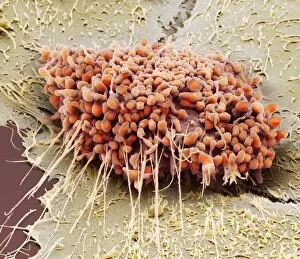
Background imageImages Dated 1st November 2012: Mouth cancer cells, SEM

Mouth cancer cells, SEM
Mouth cancer cells. Coloured scanning electron micrograph (SEM) of squamous cell carcinoma (cancer) cells from a human mouth

Background imageImages Dated 1st November 2012: Mouth cancer cells, SEM

Mouth cancer cells, SEM
Mouth cancer cells. Coloured scanning electron micrograph (SEM) of squamous cell carcinoma (cancer) cells from a human mouth

Background imageImages Dated 1st November 2012: Intestinal brush border, TEM

Intestinal brush border, TEM
Intestinal brush border. Transmission electron micrograph (TEM) of a section through a brush border (centre) in the small intestine

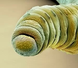
Background imageImages Dated 1st November 2012: Earthworm head, SEM

Earthworm head, SEM
Earthworm head. Coloured scanning electron micrograph (SEM) of the head of a small annelid (segmented) earthworm, showing its mouth, and small clumps of setae (bristles)

Background imageImages Dated 1st November 2012: Intestinal brush border, TEM

Intestinal brush border, TEM
Intestinal brush border. Transmission electron micrograph (TEM) of a section through a brush border (centre) in the small intestine

Background imageImages Dated 1st November 2012: Bdelloid rotifer, SEM

Bdelloid rotifer, SEM
Bdelloid rotifer. Coloured scanning electron micrograph (SEM) of a rotifer (class Bdelloidea) from a pond. Rotifers (phylum Rotifera) are microscopic aquatic animals that are related to roundworms

Background imageImages Dated 1st November 2012: ATP sulfurylase molecule

ATP sulfurylase molecule. Computer model showing the structure of an ATP sulfurylase (ATPS) enzyme. ATPS catalyses the primary step of sulphate activation within cells

Background imageImages Dated 1st November 2012: Mouth cancer cell, SEM

Mouth cancer cell, SEM
Mouth cancer cell. Coloured scanning electron micrograph (SEM) of a squamous cell carcinoma (cancer) cell from a human mouth

Background imageImages Dated 1st November 2012: Flu virus surface protein and antibody

Flu virus surface protein and antibody. Molecular model of the neuraminidase glycoprotein enzyme found on the surface of the influenza (flu) virus

Background imageImages Dated 1st November 2012: Entomophthora muscae parasitising a fly

Entomophthora muscae parasitising a fly
A bluebottle fly (Calliphora vomitoria) killed by the parasitic fungus (Entomophthora muscae). The fungus is highly adapted to its host

Background imageImages Dated 1st November 2012: Myelinated nerve, TEM

Myelinated nerve, TEM
Myelinated nerve. Transmission electron micrograph (TEM) of a section through a small myelinated nerve fibre (axon, circular, centre) from the lining of the trachea (windpipe)

Background imageImages Dated 1st November 2012: Mouth cancer cell dividing, SEM

Mouth cancer cell dividing, SEM
Mouth cancer dividing. Coloured scanning electron micrograph (SEM) of a squamous cell carcinoma (cancer) cell from a human mouth undergoing mitosis (nuclear division)

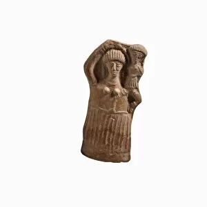
Background imageImages Dated 1st November 2012: Terra Cotta female figurine

Terra Cotta female figurine. She is depicted carrying her child on her shoulder

Background imageImages Dated 1st November 2012: Sensory rhodopsin transducer molecule

Sensory rhodopsin transducer molecule. Computer model showing the molecular structure of sensory rhodopsin transducer (SRT)

Background imageImages Dated 1st November 2012: Mouth cancer cell, SEM

Mouth cancer cell, SEM
Mouth cancer cell. Coloured scanning electron micrograph (SEM) of a squamous cell carcinoma (cancer) cell from a human mouth

Background imageImages Dated 1st November 2012: Parthian ceramics

Parthian ceramics 2nd century BCE

Background imageImages Dated 1st November 2012: HIV antibody therapy, molecular model

HIV antibody therapy, molecular model
HIV antibody therapy. Molecular model of the interaction of the HIV surface protein gp120 (green) as it interacts with a human white blood cell surface protein (CD4)

Background imageImages Dated 1st November 2012: Nebatean terracotta vessel

Nebatean terracotta vessel
A Nebatean terracotta vessel in the shape of an animal carrying two juga 1sy century BCE

Background imageImages Dated 1st November 2012: Mouth cancer cell, SEM

Mouth cancer cell, SEM
Mouth cancer cell. Coloured scanning electron micrograph (SEM) of a squamous cell carcinoma (cancer) cell from a human mouth

Background imageImages Dated 1st November 2012: Intestinal brush border, TEM

Intestinal brush border, TEM
Intestinal brush border. Transmission electron micrograph (TEM) of a section through a brush border in the small intestine

Background imageImages Dated 1st November 2012: Mouth cancer cells, SEM

Mouth cancer cells, SEM
Mouth cancer cells. Coloured scanning electron micrograph (SEM) of squamous cell carcinoma (cancer) cells from a human mouth

Background imageImages Dated 1st November 2012: Mouth cancer cell, SEM

Mouth cancer cell, SEM
Mouth cancer cell. Coloured scanning electron micrograph (SEM) of a squamous cell carcinoma (cancer) cell from a human mouth

Background imageImages Dated 1st November 2012: Tumour suppressor protein molecular model

Tumour suppressor protein molecular model
Tumour suppressor protein, molecular model. P53 prevents the proliferation of cells with damaged DNA. Its production occurs in response to radiation and chemicals that damage the structure of DNA

Background imageImages Dated 1st November 2012: Muscle arteriole, TEM

Muscle arteriole, TEM
Muscle arteriole. Transmission electron micrograph (TEM) of a section through an arteriole (white, centre) in striated muscle tissue. Magnification: x3500 when printed 10 centimetres wide

Background imageImages Dated 1st November 2012: Gene activator protein

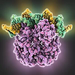
Gene activator protein. Molecular model of catabolite gene activator protein (CAP, purple) bound to a molecule of deoxyribonucleic acid (DNA, green and yellow)

Background imageImages Dated 1st November 2012: Human poliovirus, molecular model

Human poliovirus, molecular model
Human poliovirus particle. Computer model of the capsid of the human poliovirus. The capsid is a protein coat that encloses the viruss genetic information (genome), stored as RNA (ribonucleic acid)

Background imageImages Dated 1st November 2012: Mouth cancer cells, SEM

Mouth cancer cells, SEM
Mouth cancer cells. Coloured scanning electron micrograph (SEM) of squamous cell carcinoma (cancer) cells from a human mouth


All Professionally Made to Order for Quick Shipping


-

Why Choose Us?

We are a leading provider of Art Prints and Photo Gifts since 2004, working in partnership with a range of Sporting Clubs, Charities, Museums and Picture Libraries. A large share of profits from any of their images will go directly towards supporting that charity or club. Our archive of images is carefully curated to bring you a wide range of subjects, including landscapes, wildlife, architecture, and more. We ship from our partner labs in the UK, USA, EU (Netherlands) and Australia.
+

How do I place an order?

Ordering is quick & easy - Just follow 5 Simple Steps:
  1. Find your image: Use our search box or browse our online photo Collections to find the image you want.

  2. Choose your print format: Select your desired product and add it to your cart.

  3. Enter your details: If you're a returning customer, simply enter your email address and password, and we'll fill in your billing and shipping address details. All personal details are held securely and are fully GDPR compliant. As standard, we remove all Personally Identifiable Information after 12 months.

  4. Pay for your purchase: We use state-of-the-art security for online shopping and do not have access to your card details.

  5. Sit back and relax: We'll email you confirmation of your order and when it's dispatched. Registered customers can also track orders in the 'My Account' area.

+

How do I pay for an order?

You can pay for your order with most of the major credit and debit cards, or PayPal. For added security, major financial institutions process payment details separately and securely on our behalf. We do not have access to your online payment card details. Online payments are preferred however we do take phone orders. For UK customers only we accept cheques issued against a UK bank.
+

Is my data safe?

We take data security very seriously. We do not have access to your full card details and all payments are requested over a fully secure connection. Additionally, we fully comply with current European and GDPR legislation, and automatically remove all personal data after at most 12 months (unless you have an account with us and have signed in recently). We also have a strict opt-in policy and would never sell your personal details. Your data is only used to fulfill your order promptly and efficiently.
+

Is wall art ready to hang?

For quick and easy installation all wall art, including framed prints, canvas prints and metal prints are supplied with a ready to hang solution on the back. Generally, saw tooth hangers are applied as they allow wall art to hang flush against the wall. The serrated edge of the hanger prevents the frame from shifting or tilting when hung.
+

Are the photo prints fade resistant?

Yes, we use archival quality photo paper photographic paper for vivid reproduction Prints are an accurate representations of the original artwork, which is preserved for artistic character and authenticity. We guarantee they match previews shown on our web site