Fraud Blocker Skip to main content

Home > All Images > 2013 > March > 27 Mar 2013

Images Dated 27th March 2013 (page 5)

675 items

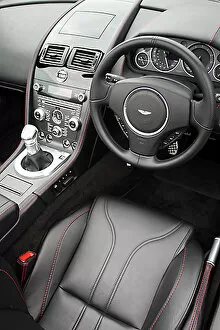
Background imageImages Dated 27th March 2013: Aston Martin V12 Vantage Roadster

Aston Martin V12 Vantage Roadster 2012 Red

Background imageImages Dated 27th March 2013: Aston Martin V12 Vantage Roadster

Aston Martin V12 Vantage Roadster 2012 Red

Background imageImages Dated 27th March 2013: Aston Martin V12 Vantage Roadster

Aston Martin V12 Vantage Roadster 2012 Red

Background imageImages Dated 27th March 2013: Aston Martin V12 Vantage Roadster

Aston Martin V12 Vantage Roadster 2012 Red

Background imageImages Dated 27th March 2013: Aston Martin V12 Vantage Roadster

Aston Martin V12 Vantage Roadster 2012 Red

Background imageImages Dated 27th March 2013: Aston Martin V12 Vantage Roadster

Aston Martin V12 Vantage Roadster 2012 Red

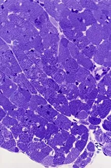
Background imageImages Dated 27th March 2013: Gastritis, light micrograph

Gastritis, light micrograph
Gastritis. Light micrograph of a section through a stomach tissue, showing chronic gastritis (inflammation). This is characterised by disorganisation of the surface mucosa lining and its glands

Background imageImages Dated 27th March 2013: Gray wolf

Gray wolf
North American timber wolves (Canis lupus) at the Wolf Science Center, Austria. The gene for melanism (dark coloured coats)

Background imageImages Dated 27th March 2013: Bone tissue, light micrograph

Bone tissue, light micrograph
Bone tissue. Darkfield illuminated light micrograph of a section through mature bone tissue, showing the concentric patterns of osteocyte cells within osteons

Background imageImages Dated 27th March 2013: Gray wolf sleeping

Gray wolf sleeping
Gray wolf (Canis lupus) sleeping at the Wolf Science Center, Austria. This research laboratory explores the similarities and links between wolves, dogs and humans

Background imageImages Dated 27th March 2013: Oocyte, light micrograph

Oocyte, light micrograph
Oocyte. Light micrograph of a section through an oocyte within a early antral follicle in an ovary. Oocytes are immature ova, or egg cells

Background imageImages Dated 27th March 2013: Sciatica, MRI scan

Sciatica, MRI scan
Sciatica. Coloured magnetic resonance imaging (MRI) scan of a sagittal section through the lumbar (lower back) spine of a 35-year-old patient

Background imageImages Dated 27th March 2013: Influenza proton pump, molecular model

Influenza proton pump, molecular model
Influenza proton pump. Molecular model showing the protein structure of a proton pump from an influenza A virus. Proton pumps are membrane proteins that move protons across a cell membrane

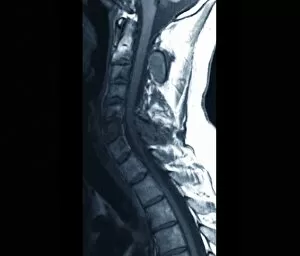
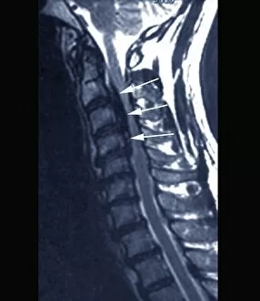
Background imageImages Dated 27th March 2013: Spinal cancer, MRI scan

Spinal cancer, MRI scan
Spinal cancer. Magnetic resonance imaging (MRI) scan of a sagittal section through the cervical (neck) region of the spine of a 22-year-old patient, showing the presence of bone lesions in the C3

Background imageImages Dated 27th March 2013: Influenza proton pump, molecular model

Influenza proton pump, molecular model
Influenza proton pump. Molecular model showing the protein structure of a proton pump from an influenza A virus. Proton pumps are membrane proteins that move protons across a cell membrane

Background imageImages Dated 27th March 2013: Cardiac muscle, light micrograph

Cardiac muscle, light micrograph
Cardiac muscle. Light micrograph of a section through muscle tissue from a heart, showing the cardiomyocyte cells. Heart muscle cells show central nuclei but the dominant feature is the abundance of

Background imageImages Dated 27th March 2013: Phospholamban membrane protein molecule

Phospholamban membrane protein molecule
Phospholamban membrane protein. Molecular model of the type II membrane protein phospholamban (PLN), which is found in cardiac muscle cells

Background imageImages Dated 27th March 2013: Spondylodiscitis, MRI scan

Spondylodiscitis, MRI scan
Spondylodiscitis. Coloured magnetic resonance imaging (MRI) scan of a sagittal section through the spine of a 69-year-old patient with spondylodiscitis at the level of the T7-T8 vertebra

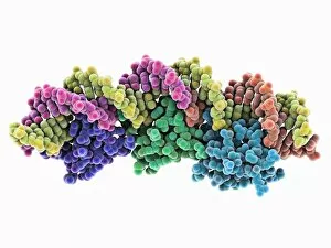
Background imageImages Dated 27th March 2013: Excisionase complex with DNA

Excisionase complex with DNA. Molecular model of three excisionase proteins (bottom, purple, green and blue) bound to a strand of DNA (top, deoxyribonucleic acid)

Background imageImages Dated 27th March 2013: Abnormal bone growth, MRI scan

Abnormal bone growth, MRI scan
Abnormal bone growth. Magnetic resonance imaging (MRI) scan of a section through the cervical spine (neck) of a 28-year-old patient, showing abnormal growth (dysplasia) of the C6 vertebra

Background imageImages Dated 27th March 2013: HIV antibody therapy, molecular model

HIV antibody therapy, molecular model
HIV antibody therapy. Molecular model of the interaction of the HIV surface protein gp120 as it interacts with a human white blood cell surface protein (CD4) and the anti-HIV antibody b12

Background imageImages Dated 27th March 2013: Gastritis, light micrograph

Gastritis, light micrograph
Gastritis. Light micrograph of a section through a stomach tissue, showing chronic gastritis (inflammation). This is characterised by disorganisation of the surface mucosa lining and its glands

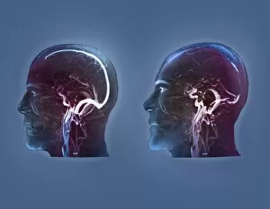
Background imageImages Dated 27th March 2013: Thrombophlebitis in the brain, MRI scans

Thrombophlebitis in the brain, MRI scans
Thrombophlebitis in the brain. Coloured magnetic resonance imaging (MRI) angiograms comparing the blood vessels (white) of the brain and neck in a healthy 43-year-old (left)

Background imageImages Dated 27th March 2013: Gastritis, light micrograph

Gastritis, light micrograph
Gastritis. Light micrograph of a section through a stomach tissue, showing chronic gastritis (inflammation). This is characterised by disorganisation of the surface mucosa lining and its glands

Background imageImages Dated 27th March 2013: Spinal cancer, MRI scan

Spinal cancer, MRI scan
Spinal cancer. Coloured magnetic resonance imaging (MRI) scan of a sagittal section through the cervical (neck) region of the spine of a 22-year-old patient

Background imageImages Dated 27th March 2013: DNA translocase, molecular model

DNA translocase, molecular model
ftsk, , protein, biomolecule, macromolecule, translocase, enzyme, pseudomonas aeruginosa, bacteria, biochemistry, biology, genetics, molecular biology, proteomics, artwork, illustration

Background imageImages Dated 27th March 2013: Colitis, light micrograph

Colitis, light micrograph
Colitis. Light micrograph of a section through colon (large intestine) tissue affected by bacterial infection, causing injury to the mucosal lining, which has resulted in inflammation (colitis)

Background imageImages Dated 27th March 2013: Gray wolf

Gray wolf (Canis lupus) at the Wolf Science Center, Austria. This research laboratory explores the similarities and links between wolves, dogs and humans

Background imageImages Dated 27th March 2013: Cardiac muscle, light micrograph

Cardiac muscle, light micrograph
Cardiac muscle. Light micrograph of a section through muscle tissue from a heart, showing the cardiomyocyte cells. Heart muscle cells show central nuclei but the dominant feature is the abundance of

Background imageImages Dated 27th March 2013: Hairy scalp skin, light micrograph

Hairy scalp skin, light micrograph
Hairy scalp skin. Light micrograph of a section through healthy skin from a human scalp, showing the numerous hair follicles. The scalp is an example of hairy skin

Background imageImages Dated 27th March 2013: Oxalosis, light micrograph

Oxalosis, light micrograph
Oxalosis. Light micrograph of a section through adult bone tissue affected by oxalosis (primary hyperoxaluria), showing mature bone (green)

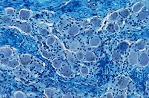
Background imageImages Dated 27th March 2013: Peripheral nerve, light micrograph

Peripheral nerve, light micrograph
Peripheral nerve. Light micrograph of a section through a peripheral nerve. This is a mixed nerve with myelinated axons (dark blue circles)

Background imageImages Dated 27th March 2013: Ischaemic bowel, light micrograph

Ischaemic bowel, light micrograph
Ischaemic bowel. Light micrograph of a section through a portion of intestine that has been damaged by an interruption of the blood supply, or ischaemia

Background imageImages Dated 27th March 2013: Nerve ganglion, light micrograph

Nerve ganglion, light micrograph
Nerve ganglion. Light micrograph of a section through a dorsal (sensory) spinal root ganglion associated with a sensory nerve root of the spinal cord. Sensory information from peripheral sites e.g

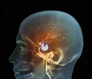
Background imageImages Dated 27th March 2013: Brain aneurysm, DSA scan

Brain aneurysm, DSA scan
Brain aneurysm. Coloured digital subtraction angiogram (DSA) of the head of a 38-year-old, showing a large aneurysm (bright, centre) of the right internal carotid artery

Background imageImages Dated 27th March 2013: Myelinated nerve, light micrograph

Myelinated nerve, light micrograph
Myelinated nerve. Light micrograph of a section through a peripheral myelinated nerve, showing many individual axons each covered with deep blue-staining myelin sheaths

Background imageImages Dated 27th March 2013: Gray wolf

Gray wolf
Wolves (Canis lupus) interacting at the Wolf Science Center, Austria. This research laboratory explores the similarities and links between wolves, dogs and humans

Background imageImages Dated 27th March 2013: Stroke, CT scan

Stroke, CT scan
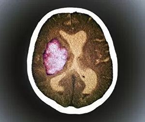
Stroke. Coloured computed tomography (CT) scan of a section through the brain of a patient 24 hours after a haemorrhagic stroke, showing an accumulation of blood (bright, centre left) in the brain

Background imageImages Dated 27th March 2013: Forestiers disease, MRI scan

Forestiers disease, MRI scan
Forestiers disease. Magnetic resonance imaging (MRI) scan of a section through the cervical spine (neck) of a 42-year-old Asian patient suffering from a stiff neck and neck pain

Background imageImages Dated 27th March 2013: DNA translocase, molecular model

DNA translocase, molecular model
ftsk, , protein, biomolecule, macromolecule, translocase, enzyme, pseudomonas aeruginosa, bacteria, biochemistry, biology, genetics, molecular biology, proteomics, artwork, illustration

Background imageImages Dated 27th March 2013: Incontinence implant, X-ray

Incontinence implant, X-ray
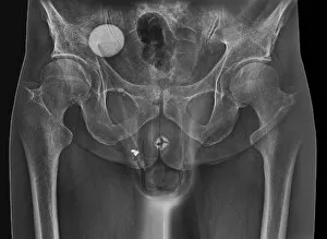
Incontinence implant. Frontal X-ray of the pelvic region of a 62-year-old patient with an artificial urinary sphincter (white, circular, lower centre) implant to control urinary incontinence

Background imageImages Dated 27th March 2013: Spina bifida, MRI scan

Spina bifida, MRI scan
Fibrolipoma of the spine. Magnetic resonance imaging (MRI) scan of a sagittal section through the spine of a 24-month-old patient with spina bifida

Background imageImages Dated 27th March 2013: Thrombophlebitis in the brain, MRI scans

Thrombophlebitis in the brain, MRI scans
Thrombophlebitis in the brain. Coloured magnetic resonance imaging (MRI) angiograms comparing the blood vessels (white) of the brain and neck in a healthy 43-year-old (left)

Background imageImages Dated 27th March 2013: Kinesin motor protein

Kinesin motor protein. Molecular model of the ncd kinesin motor protein. Kinesin motor proteins transport vesicles containing intracellular cargo around the cell along microtubules

Background imageImages Dated 27th March 2013: Incontinence implant, X-ray

Incontinence implant, X-ray
Incontinence implant. Coloured frontal X-ray of the pelvic region of a 62-year-old patient with an artificial urinary sphincter (white, circular)

Background imageImages Dated 27th March 2013: Glial cells, light micrograph

Glial cells, light micrograph
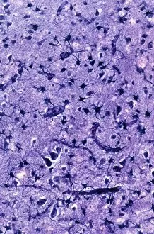
Glial cells in the brain. Light micrograph of a section through glial cells (dark) in the grey matter of the brain. Due to their star shape these glial cells are called astrocytes

Background imageImages Dated 27th March 2013: Copper sulphate crystals light micrograph

Copper sulphate crystals light micrograph
Copper sulphate crystals, polarised light micrograph. Magnification: x2.5, when printed 10 centimetres wide

Background imageImages Dated 27th March 2013: Incontinence implant, X-ray

Incontinence implant, X-ray
Incontinence implant. Coloured frontal X-ray of the pelvic region of a 62-year-old patient with an artificial urinary sphincter (white, circular)


All Professionally Made to Order for Quick Shipping


-

Why Choose Us?

We are a leading provider of Art Prints and Photo Gifts since 2003, working in partnership with a range of Sporting Clubs, Charities, Museums and Picture Libraries. A large share of profits from any of their images will go directly towards supporting that charity or club. Our archive of images is carefully curated to bring you a wide range of subjects, including landscapes, wildlife, architecture, and more. We ship from our partner labs in the UK, USA, EU (Netherlands) and Australia.
+

How do I place an order?

Ordering is quick & easy - Just follow 5 Simple Steps:
  1. Find your image: Use our search box or browse our online photo Collections to find the image you want.

  2. Choose your print format: Select your desired product and add it to your cart.

  3. Enter your details: If you're a returning customer, simply enter your email address and password, and we'll fill in your billing and shipping address details. All personal details are held securely and are fully GDPR compliant. As standard, we remove all Personally Identifiable Information after 12 months.

  4. Pay for your purchase: We use state-of-the-art security for online shopping and do not have access to your card details.

  5. Sit back and relax: We'll email you confirmation of your order and when it's dispatched. Registered customers can also track orders in the 'My Account' area.

+

How do I pay for an order?

You can pay for your order with most of the major credit and debit cards, or PayPal. For added security, major financial institutions process payment details separately and securely on our behalf. We do not have access to your online payment card details. Online payments are preferred however we do take phone orders. For UK customers only we accept cheques issued against a UK bank.
+

Is my data safe?

We take data security very seriously. We do not have access to your full card details and all payments are requested over a fully secure connection. Additionally, we fully comply with current European and GDPR legislation, and automatically remove all personal data after at most 12 months (unless you have an account with us and have signed in recently). We also have a strict opt-in policy and would never sell your personal details. Your data is only used to fulfill your order promptly and efficiently.
+

Is wall art ready to hang?

For quick and easy installation all wall art, including framed prints, canvas prints and metal prints are supplied with a ready to hang solution on the back. Generally, saw tooth hangers are applied as they allow wall art to hang flush against the wall. The serrated edge of the hanger prevents the frame from shifting or tilting when hung.
+

Are the photo prints fade resistant?

Yes, we use archival quality photo paper photographic paper for vivid reproduction Prints are an accurate representations of the original artwork, which is preserved for artistic character and authenticity. We guarantee they match previews shown on our web site