Home > Animals > Mammals > Cricetidae > White-footed Mouse
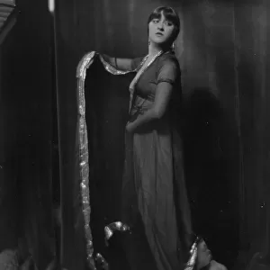
Leg and foot muscles - 1866 anatomical illustration showing the muscles (brown) and tendons (white) of the human foot (left) and leg (right) - The leg and the foot - Anatomy - Fig 1: Internal leg region- Fig 2: Dorsale region of the foot, 1866
 and tendons (white) of the human foot (left) and leg (right) - The leg and the foot - Anatomy - Fig 1: Internal leg region- Fig 2: Dorsale region of the foot, 1866")
![]()
Wall Art and Photo Gifts from Fine Art Finder
Leg and foot muscles - 1866 anatomical illustration showing the muscles (brown) and tendons (white) of the human foot (left) and leg (right) - The leg and the foot - Anatomy - Fig 1: Internal leg region- Fig 2: Dorsale region of the foot, 1866
JAB4119395 Leg and foot muscles - 1866 anatomical illustration showing the muscles (brown) and tendons (white) of the human foot (left) and leg (right) - The leg and the foot - Anatomy - Fig 1: Internal leg region- Fig 2: Dorsale region of the foot, 1866; (add.info.: Leg and foot muscles - 1866 anatomical illustration showing the muscles (brown) and tendons (white) of the human foot (left) and leg (right) - The leg and the foot - Anatomy - Fig 1: Internal leg region- Fig 2: Dorsale region of the foot, 1866); Photo ©Jaime Abecasis
Media ID 38146832
© Photo ©Jaime Abecasis / Bridgeman Images
Abecasis Anatomical Board Anatomist Foot Internal Anatomy Internal Organ Muscle
FEATURES IN THESE COLLECTIONS
> Animals
> Mammals
> Cricetidae
> White-footed Mouse
> Fine Art Finder
> Artists
> Joseph Porphyre Pinchon
EDITORS COMMENTS
This stunning 1866 anatomical illustration captures the intricate details of the leg and foot muscles, showcasing the brown muscles and white tendons in exquisite detail. The left side of the print focuses on the internal leg region, highlighting the complexity of muscle structure within our lower limbs. On the right side, we are presented with a detailed view of the dorsale region of the foot, emphasizing how these muscles and tendons work together to support our bodies.
Created during a time when anatomical knowledge was rapidly expanding, this print serves as a testament to both scientific discovery and artistic skill. The precision with which each muscle and tendon is depicted reflects not only an understanding of human anatomy but also a deep appreciation for its beauty.
As we gaze upon this historical illustration, we are reminded of the incredible complexity that lies beneath our skin. From doctors studying internal organs to artists capturing their essence on paper, this image speaks to a shared fascination with what makes us human. Let us marvel at the wonders of our own bodies and continue to explore all that they have to offer.
MADE IN AUSTRALIA
Safe Shipping with 30 Day Money Back Guarantee
FREE PERSONALISATION*
We are proud to offer a range of customisation features including Personalised Captions, Color Filters and Picture Zoom Tools
SECURE PAYMENTS
We happily accept a wide range of payment options so you can pay for the things you need in the way that is most convenient for you
* Options may vary by product and licensing agreement. Zoomed Pictures can be adjusted in the Cart.