
Adult Patient Collection
"Exploring the Complexities of an Adult Patient's Health: From Osteoarthritis to Lung Abscess" In this comprehensive medical journey


All Professionally Made to Order for Quick Shipping
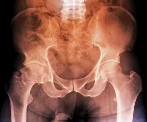
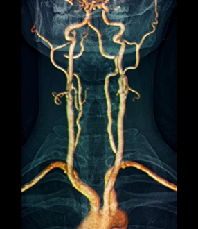
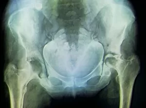
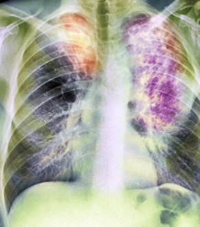
"Exploring the Complexities of an Adult Patient's Health: From Osteoarthritis to Lung Abscess" In this comprehensive medical journey, we delve into the intricate health challenges faced by an adult patient. Starting with osteoarthritis of the hip, a condition that affects mobility and causes discomfort, we turn to X-ray images that reveal the extent of damage. Moving on, calcified cysts of the ovary come into focus – mysterious formations requiring careful examination. The exploration continues as we shift our attention to 3D MRI scans capturing neck arteries supplying blood to the brain. These detailed images provide crucial insights into potential risks and help guide treatment decisions. Shifting gears again, healthy spine X-rays showcase a strong foundation while highlighting areas where fusion of spinal bones has occurred. This remarkable process is captured through multiple X-ray images, revealing how these bones have grown together over time. Transitioning from spine to pelvis, a 3D CT scan displays a vibrant image of a healthy pelvic region. This visual confirmation offers reassurance amidst other health concerns. The narrative takes another twist as lung abscesses enter center stage – detected first through X-ray imaging and then further examined using CT scans for more precise evaluation. These abscesses represent pockets of infection within lung tissue that require immediate attention and targeted treatment plans. Lastly, atelectasis or lung collapse becomes evident in yet another X-ray image - emphasizing the importance of prompt intervention in restoring normal lung function. Through this captivating journey inside an adult patient's complex health landscape, medical professionals gain invaluable knowledge necessary for diagnosis and tailored treatments.