Fraud Blocker Skip to main content

Algal Collection (page 4)

"Exploring the Intricate World Beauty" Immerse yourself in the captivating artistry of diatom algae, as depicted by Ernst Haeckel

Background imageAlgal Collection: Netrium desmids, light micrograph

Netrium desmids, light micrograph
Netrium desmids. Polarised light micrograph of Netrium sp. desmid green alga. Desmids are a common group of freshwater single-celled algae that have intricate cell walls

Background imageAlgal Collection: Cosmarium desmids, light micrograph

Cosmarium desmids, light micrograph
Phase contrast, magnification: 200 X on APS-C sensor

Background imageAlgal Collection: Euastrum desmids, light micrograph

Euastrum desmids, light micrograph
Euastrum desmids. Polarised light micrograph of Euastrum sp. desmid green algae. Desmids are a common group of freshwater single-celled algae that have intricate cell walls

Background imageAlgal Collection: Micrasterias desmids, light micrograph

Micrasterias desmids, light micrograph
Micrasterias desmids. Polarised light micrograph of Micrasterias truncata desmid green algae. Desmids are a common group of freshwater single-celled algae that have intricate cell walls
Background imageAlgal Collection: Micrasterias desmid, light micrograph

Micrasterias desmid, light micrograph
Micrasterias desmid. Differential interference contrast light micrograph of a Micrasterias sp. desmid green alga. Desmids are a common group of freshwater single-celled algae that have intricate cell

Background imageAlgal Collection: Plankton and fish, artwork C016 / 3448

Plankton and fish, artwork C016 / 3448
Plankton and fish. Artwork of plankton as viewed under a magnifying glass, with seabream (Diplodus vulgaris) fish

Background imageAlgal Collection: Blue green algae stigonema, LM C013 / 5286

Blue green algae stigonema, LM C013 / 5286
Light micrograph using differential interference contrast microscopy (DIC) of the filamentous blue-green algae (group name Cyanophycophyta), called stigonema

Background imageAlgal Collection: LM of the red algae, Plumaria elegans C013 / 5279

LM of the red algae, Plumaria elegans C013 / 5279
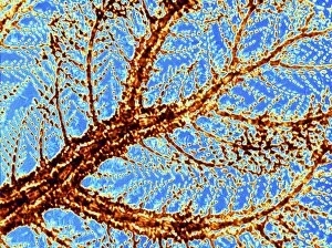
Plumaria algae. False color light micrograph of part of the finely branched thallus of the red algae Plumaria elegans (family: Florideophycidae)

Background imageAlgal Collection: Red algae, light micrograph C013 / 5281

Red algae, light micrograph C013 / 5281
Red algae. Light micrograph of filaments of the red alga Polysiphonia sp. This marine algae is found throughout the world inhabiting rock pools and water up to 27 metres deep

Background imageAlgal Collection: Diploneis diatom, SEM C013 / 5110

Diploneis diatom, SEM C013 / 5110
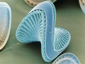
Diploneis diatom, coloured scanning electron micrograph (SEM). Diatoms are single-celled photosynthetic algae, of which there are about 100, 000 species

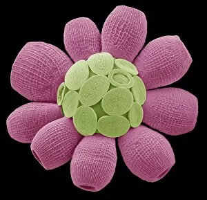
Background imageAlgal Collection: Coccolithophorid plankton, SEM C013 / 5109

Coccolithophorid plankton, SEM C013 / 5109
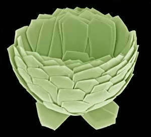
Coccolithophorid plankton. Coloured scanning electron micrograph (SEM) of the external mineralised structure (coccosphere) of a small marine algal organism called a coccolithophore

Background imageAlgal Collection: LM of the colonial diatom Fragillaria sp

LM of the colonial diatom Fragillaria sp
Light micrograph of the diatom Fragillaria sp.. The filaments seen here are simple colonies of Fragillaria. They consist of a stack of diatoms which have failed to separate following cell division

Background imageAlgal Collection: Colonial alga, Dinobryon

Colonial alga, Dinobryon
Light micrograph of a branching colony of the golden- brown alga Dinobryon sp.. The colony is composed of single-celled algae, each of which secretes a fine, transparent

Background imageAlgal Collection: Melosira filamentous diatom alage, LM

Melosira filamentous diatom alage, LM
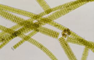
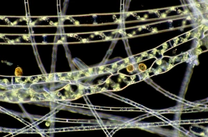
Melosira sp. diatoms. Light micrograph of filamentous strands of the freshwater diatom alga Melosira sp.. Diatoms are single-celled algae that form cell walls made of silica

Background imageAlgal Collection: SEM of blue-green algae

SEM of blue-green algae
Blue-green algae. Coloured scanning electron micrograph (SEM) of Oscillatoria sp. filamentous blue-green algae. Blue-green algae are considered to be one of the most primitive organisms

Background imageAlgal Collection: Oscillatoria blue-green alga

Oscillatoria blue-green alga
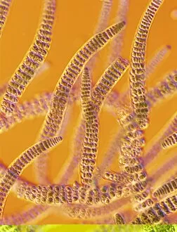
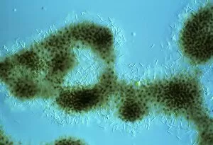
Light micrograph of Oscillatoria sp. (brown strands), a filamentous blue-green alga or cyanobacterium. The strands of alga are in fact linear colonies of primitive, single-celled

Background imageAlgal Collection: Diatom algae, Campylodiscus

Diatom algae, Campylodiscus
Diatoms. Coloured scanning electron micrograph of the diatom Campylodiscus hibernicus. The diatoms are a distinctive group of single-celled algae containing about 10, 000 species

Background imageAlgal Collection: Colonial freshwater diatoms

Colonial freshwater diatoms
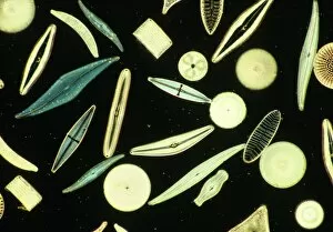
Light micrograph of assorted freshwater diatoms. The rectangular, broken filaments seen here are simple colonies. They consist of a chain of diatoms which have failed to separate following cell

Background imageAlgal Collection: Light micrograph of assorted diatoms

Light micrograph of assorted diatoms. The diatoms are a distinctive group of single-celled algae containing about 10, 000 species

Background imageAlgal Collection: Chlamydomonas sp. algae, SEM

Chlamydomonas sp. algae, SEM
Green algae. Coloured scanning electron micrograph (SEM) of two Chlamydomonas sp. green algae. These single-celled organisms are aquatic, living in freshwater habitats

Background imageAlgal Collection: Green alga, Micrasterias

Green alga, Micrasterias
Micrasterias alga. Coloured scanning electron micrograph (SEM) of the desmid Micrasterias sp.. The desmids are a group of freshwater unicellular green algae characterised by their intricately- shaped

Background imageAlgal Collection: Conjugating spirogyra

Conjugating spirogyra
Light micrograph showing conjugating filaments of Spirogyra, a green alga which forms slimy threads in stagnant or slow moving water

Background imageAlgal Collection: LM of Chlorella surrounded by bacteria

LM of Chlorella surrounded by bacteria
Light micrograph of Chlorella sp. a unicellular green alga which inhabits freshwater ponds and lakes. The non-motile, globular cells may be solitary or aggregated into groups as seen here

Background imageAlgal Collection: LM of conjugating filaments of Spirogyra

LM of conjugating filaments of Spirogyra
Light micrograph showing conjugating filaments of Spirogyra sp. a green alga which forms slimy threads in stagnant or slow moving water

Background imageAlgal Collection: Filaments of Spirogyra alga

Filaments of Spirogyra alga
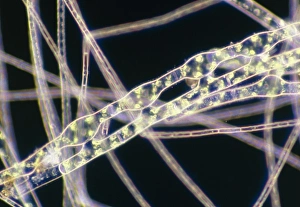
Light micrograph of filaments of Spirogyra, a green alga which forms slimy threads called water- silk" or " mermaids tresses" in stagnant or slow moving water

Background imageAlgal Collection: Scyphosphaera apsteinii

Scyphosphaera apsteinii. SEM image of an equatorial coccolith

Background imageAlgal Collection: Model of coccolith Kamptnerius magnificus

Model of coccolith Kamptnerius magnificus
Copy of the resin model of the coccolith Kamptnerius magnificus Deflandre, 1930 currently on display in the Earth Lab area of the Earth Galleries at the Natural History Museum, London

Background imageAlgal Collection: Usnea inflata, beard lichen

Usnea inflata, beard lichen
And Hypogymnia physodes (right), Burnham Beeches, Bucks, W. London. The former is a recent colonist following reductions in SO2 pollution

Background imageAlgal Collection: Ceratolithoides aculeus, coccolith

Ceratolithoides aculeus, coccolith
Scanning electron microscope image of an isolated coocolith from Cretaceous chalk. These are thin calcite shells protecting the coccolithophore within

Background imageAlgal Collection: Chorda filum, sea lace

Chorda filum, sea lace
Cut out of mounted specimen of sea lace or Dead mans rope. A brown seaweed, this specimen is 14.5 feet long and held at the Natural History Museum, London

Background imageAlgal Collection: Florosphaera profunda, coccolith

Florosphaera profunda, coccolith
Scanning electron microscope image of a complete sphere of coccoliths from modern oceans. These are thin calcite shells protecting the coccolithophore within

Background imageAlgal Collection: Proterozoic ocean floor

Proterozoic ocean floor
A restoration of Proterozoic ocean floor with bun-shaped stromatolites

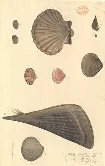
Background imageAlgal Collection: Nine molluscs, including bivalves and gastropods

Nine molluscs, including bivalves and gastropods
Watercolour 391 by the Port Jackson Painter, entitled Kow-er-ring, Kow-ill, Kaa-din, Wal-gan, from the Watling Collection

Background imageAlgal Collection: Dumontia contorta, seaweed

Dumontia contorta, seaweed

Background imageAlgal Collection: Scinaia forcellata, seaweed

Scinaia forcellata, seaweed
Cut out of specimen of marine alga or seaweed collected by Holmes in Enoura, Japan. Specimen is held in the Crypt. Herbarium at the Natural History Museum, London

Background imageAlgal Collection: Emiliania huxleyi coccosphere

Emiliania huxleyi coccosphere
Coccosphere of Emiliania huxleyi from the Western Mediterranean. E. huxleyi is one of the most widespread species on earth

Background imageAlgal Collection: Desmarestia ligulata, seaweed

Desmarestia ligulata, seaweed
Page 55 from Algae Danmonienses: or dried specimens of Marine Plants, principally collected in Devonshire by Mary Wyatt; carefully named according to Dr. Hookers British Flora

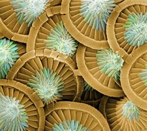
Background imageAlgal Collection: Amphitetras, diatom

Amphitetras, diatom
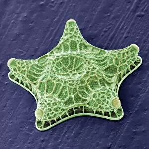
Scanning electron microscope (SEM) image showing the diatom Amphitetras with its ornate silica shell (x5000 on a standard 9 cm wide print). Coloured artificially by computer

Background imageAlgal Collection: Frontispiece of Catalogue Raisonne d une collection

Frontispiece of Catalogue Raisonne d une collection
Illustration by Francois Boucher from the book Catalogue Raisonne d une collection by Edme Francois Gersaint, 1744

Background imageAlgal Collection: The pond in the Wildlife Garden

The pond in the Wildlife Garden. Photographed by Derek Adams. Published in Wildlife Garden by Roy Vickery, 2004 page 35

Background imageAlgal Collection: Ciliate plankton

Ciliate plankton
Scanning electron microscope image of a ciliate showing clearly the microscopic hairs or cilia that they use for movement and feeding (x 700)

Background imageAlgal Collection: Fucus vesiculosis, bladderwrack

Fucus vesiculosis, bladderwrack
Illustration from Botany Library Plate Collection at the Natural History Museum, London. By Leopald Trattinick, 1825

Background imageAlgal Collection: Coccoliths

Coccoliths
Scanning electron microscope (SEM) image of coccoliths, these are the limestone scales surrounding the marine phytoplankton coccolithophores

Background imageAlgal Collection: Acanthoica acanthifera

Acanthoica acanthifera

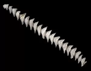
Background imageAlgal Collection: Spiral axis of Archimedes, bryozoan

Spiral axis of Archimedes, bryozoan
Archimedes, was a bryozoan possibly living in association with an alga. From the Lower Carboniferous limestone, Iowa, USA. c. 350-330 million years old

Background imageAlgal Collection: Coccolithus pelagicus

Coccolithus pelagicus
Coccosphere of Coccolithus pelagicus, a common cold water coccolithophore. Collected from the British Continental shelf, North West of Scotland. Specimen diameter 15m. False-coloured SEM image

Background imageAlgal Collection: Florisphaera profunda

Florisphaera profunda
A coccolithophore with highly modified, plate-like coccoliths. This is a very common deep dwelleing species, typically living at about 100-150m depth in the water column


All Professionally Made to Order for Quick Shipping

"Exploring the Intricate World Beauty" Immerse yourself in the captivating artistry of diatom algae, as depicted by Ernst Haeckel. These microscopic organisms showcase their intricate structures and mesmerizing patterns, reminding us of the boundless creativity found in nature. Pressed seaweed specimens C016 / 6127 offer a glimpse into the diverse forms and textures that algal species can take. From delicate filaments to robust fronds, each specimen tells a unique story of adaptation and survival in marine ecosystems. Behold the beauty of calcareous phytoplankton through scanning electron microscopy (SEM). The exquisite details captured reveal their ornate shells and elaborate architecture, serving as a testament to their vital role in oceanic food webs. Discosphaera tubifera, a coccolithophore, enchants with its spherical shape adorned by intricate calcium carbonate plates. This tiny organism plays an essential part in carbon cycling and contributes to the stunning white cliffs seen along some coastlines. Dictyota dichotoma showcases its elegant branching structure, exemplifying how algae can create complex habitats for other marine organisms. Its presence enriches coastal ecosystems while providing shelter for countless creatures. Diatoms take center stage once again under SEM's magnifying lens. Their symmetrical siliceous skeletons exhibit remarkable diversity – from star-shaped patterns to delicate lace-like designs – showcasing nature's endless ingenuity. Marvel at Fucus bulbosus or kelp's majestic presence as it sways gracefully beneath ocean currents. These large brown algae provide refuge for numerous marine species while contributing to nutrient cycling within coastal environments. Microcystis blue-green alga captivates with its vibrant hue amidst freshwater bodies. Though often associated with harmful algal blooms, this organism also serves ecological functions such as nitrogen fixation and oxygen production. Witness yet another diatom masterpiece through SEM imagery – their intricately sculpted shapes and delicate ornamentation never cease to amaze.