
Alveolar Collection
"Exploring the Intricacies of Alveolar: A Journey through Art and Anatomy" Step into a world where art meets science, as we delve into the captivating realm of alveolar


All Professionally Made to Order for Quick Shipping
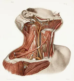
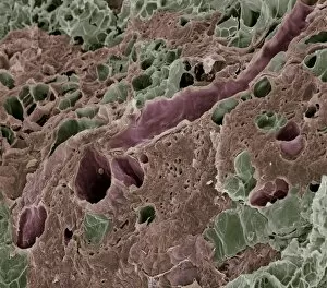
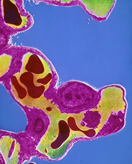
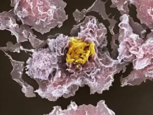
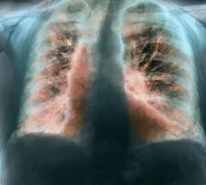
"Exploring the Intricacies of Alveolar: A Journey through Art and Anatomy" Step into a world where art meets science, as we delve into the captivating realm of alveolar. From neck vascular anatomy to historical artwork, this 150-word caption will take you on an enlightening journey. Let's start with a glimpse into the intricate network of neck vascular anatomy. Marvel at the detailed depictions in historical artwork that showcase the delicate balance between form and function. Now, shift your focus to lungs anatomy – a fascinating landscape filled with tiny structures called alveoli. Explore breathtaking artwork that captures their beauty, showcasing their role in oxygen exchange within our bodies. But there's more to alveolar than just its presence in lung tissue. Witness lactating breast tissue under scanning electron microscopy (SEM), revealing its unique structure and function during breastfeeding. Delving deeper, observe capillaries within the alveolar septum of lungs through transmission electron microscopy (TEM). These images provide a closer look at these vital blood vessels' architecture and their crucial role in gas exchange. However, not all is well within these microscopic worlds. Observe TB bacteria infecting macrophages under SEM – a stark reminder of the challenges faced by our immune system against infectious diseases like tuberculosis. Lastly, witness fibrosing alveolitis captured through X-ray imaging. This condition showcases how inflammation can lead to scarring and thickening of lung tissues, impacting respiratory health. From artistry to scientific exploration, this captivating journey has shed light on various aspects intricacies. It reminds us that even within seemingly small structures lies immense complexity waiting to be discovered and understood for both artistic inspiration and medical advancement.