
Anatomical Model Collection
"Exploring the Intricacies of the Human Body: Anatomical Models Unveiled" Delving into the wonders of human anatomy


All Professionally Made to Order for Quick Shipping
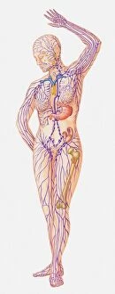
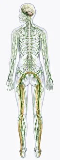
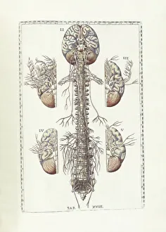
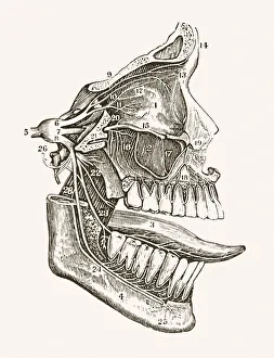
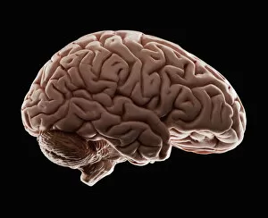
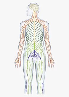
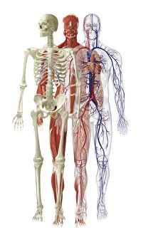
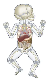
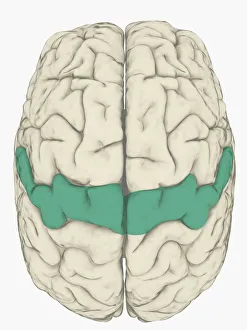
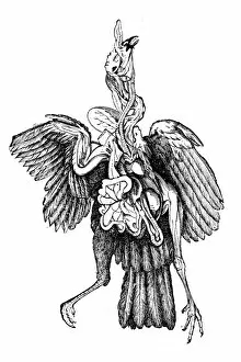
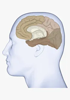
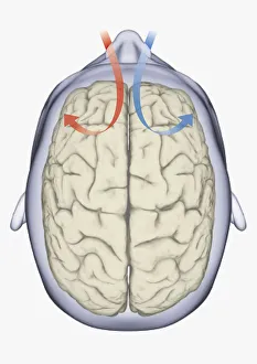
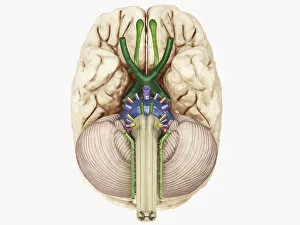
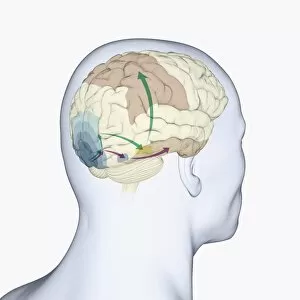
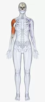
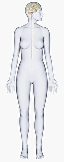
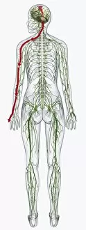
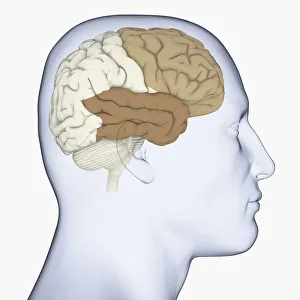
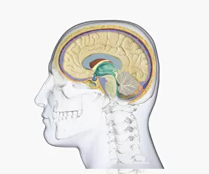
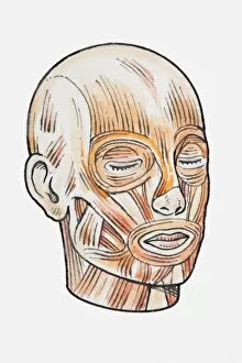
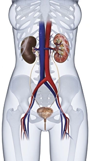
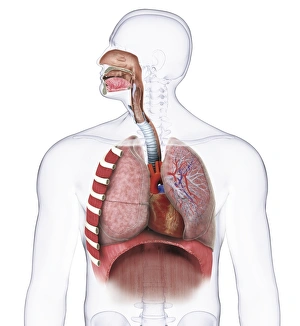
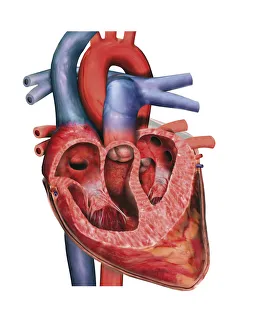
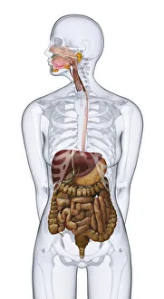
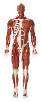
"Exploring the Intricacies of the Human Body: Anatomical Models Unveiled" Delving into the wonders of human anatomy, these anatomical models provide a captivating insight into our complex systems. From an exquisite illustration showcasing the female lymphatic system, to a digital representation revealing the intricate connections between the nervous system and spinal cord, each model offers a unique perspective on our inner workings. Transporting us back in time, Bartholomeo Eustachi's masterpiece "The Science of Human Anatomy" takes center stage with its meticulous depiction of bodily structures. Meanwhile, a 19th-century medical illustration unveils the enigmatic network of facial nerves that have fascinated scientists for centuries. A studio-shot model captures our attention as it showcases the intricacies of the human brain. With spinal nerves branching out from its core, this digital illustration highlights how every part plays a vital role in maintaining our well-being. Moving beyond just one region, another digital illustration reveals muscles spanning across necks, arms, chests, diaphragms, and upper legs - emphasizing their importance in movement and stability. Zooming closer to specific brain functions brings forth intriguing discoveries. A profile view illustrates somatosensory cortex highlighted in pink while another highlights it in blue - unraveling its significance in sensory perception. Diving deeper into neurology leads us to explore areas such as mirror neurons within our brains. Through an orange hue illuminating specific regions within a head profile view; we gain insights into empathy and understanding others' actions. Not limited solely to humans but extending even further; turkey anatomy is also unveiled through detailed illustrations - reminding us that comparative studies can broaden our knowledge base. These anatomical models serve as windows into understanding ourselves better; they remind us of both complexity and beauty intertwined within each individual's body.