Fraud Blocker Skip to main content

Anatomy Collection (page 100)

"Unveiling the Intricacies of Anatomy: From Sensory Homunculus to Motor Homunculus" Step into the fascinating world of anatomy, where every detail tells a story

Background imageAnatomy Collection: Human speech organs, wood engraving, published in 1880

Human speech organs, wood engraving, published in 1880
The Organs of Speech: A) mouth, B) uvula, C) tongue, D) tonsils, E) epiglottis, F) thyroid cartilage, G) arytenoid muscle, H+I) vestibular folds, K) vocal fold, L) larynx, M) hypopharynx, N) trachea

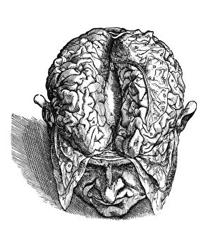
Background imageAnatomy Collection: The Human Brain

The Human Brain
circa 1870: Anatomical drawing of a mans brain and cerebral nerves. (Photo by Hulton Archive/Getty Images)

Background imageAnatomy Collection: Picture No. 10873599

Picture No. 10873599
Human Crab Louse (Phthirus pubis) Date:

Background imageAnatomy Collection: Picture No. 10851731

Picture No. 10851731
Norwegian Lundehund Dog - Foot Date:

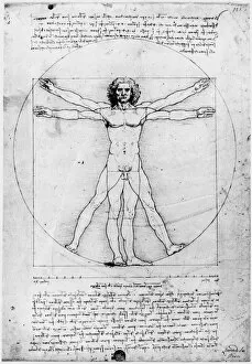
Background imageAnatomy Collection: Proportions of the Human Figure or The Vitruvian Man. Pen and ink drawing by Leonardo Da Vinci

Proportions of the Human Figure or The Vitruvian Man. Pen and ink drawing by Leonardo Da Vinci, c1492
LEONARDO: ANATOMY. Proportions of the Human Figure or The Vitruvian Man. Pen and ink drawing by Leonardo Da Vinci, c1492

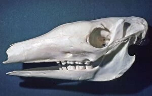
Background imageAnatomy Collection: Picture No. 10874838

Picture No. 10874838
Pigmy Hippo skull (Hexaprotodon liberiensis) Date:

Background imageAnatomy Collection: Picture No. 10865930

Picture No. 10865930
Bamboo - cross section of a shoot emerging from the ground and its rhizome Date:

Background imageAnatomy Collection: Picture No. 10864939

Picture No. 10864939
Mink Skull- X ray showing teeth (Mustela vison) Date:

Background imageAnatomy Collection: Picture No. 10864934

Picture No. 10864934
Aardvark - skull showing simple peg like cheek teeth and no incisors or canines (Orycteropus afer) Date:

Background imageAnatomy Collection: Picture No. 10861874

Picture No. 10861874
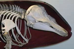
Harbour Porpoise - Skeleton inside shape of skin (Phocoena phocoena) Date:

Background imageAnatomy Collection: Picture No. 10854313

Picture No. 10854313
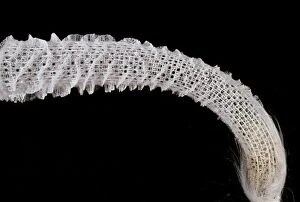
Venus Glass Sponge / Flower Basket (Euplectella) Date:

Background imageAnatomy Collection: Picture No. 10852952

Picture No. 10852952
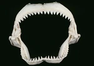
Shark Jaws - skeleton (Carcharhinus sp.) Date:

Background imageAnatomy Collection: Picture No. 10851777

Picture No. 10851777
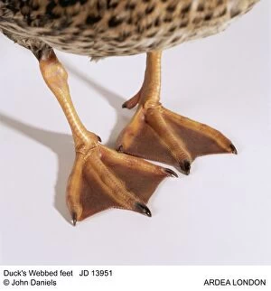
Duck's Webbed feet Date:

Background imageAnatomy Collection: Picture No. 10745632

Picture No. 10745632
Horse. Split hoof Date:

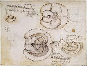
Background imageAnatomy Collection: Pen and ink drawing, c1508, by Leonardo da Vinci of the cerebral hemispheres and ventricles

Pen and ink drawing, c1508, by Leonardo da Vinci of the cerebral hemispheres and ventricles
LEONARDO: VENTRICLES, c1508. Pen and ink drawing, c1508, by Leonardo da Vinci of the cerebral hemispheres and ventricles

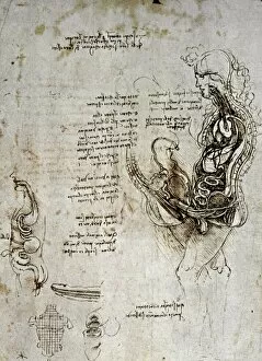
Background imageAnatomy Collection: Drawing of the human sexual organs

Drawing of the human sexual organs
LEONARDO DA VINCI. Drawing of the human sexual organs

Background imageAnatomy Collection: Chinese acupuncture chart showing the kidney meridian

Chinese acupuncture chart showing the kidney meridian
ACUPUNCTURE CHART. Chinese acupuncture chart showing the kidney meridian

Background imageAnatomy Collection: Dissection of the brain (fig. 3). Woodcut from the seventh book of Andreas Vesalius De Humani

Dissection of the brain (fig. 3). Woodcut from the seventh book of Andreas Vesalius De Humani Corporis Fabrica
VESALIUS: BRAIN, 1543. Dissection of the brain (fig.3). Woodcut from the seventh book of Andreas Vesalius " De Humani Corporis Fabrica, " published in 1543 at Basel

Background imageAnatomy Collection: Anatomical / Astrological Man. Miniature depicting the influence of the zodiacal stars

Anatomical / Astrological Man. Miniature depicting the influence of the zodiacal stars on the human body from the 15th
ANATOMICAL MAN. Anatomical/Astrological Man. Miniature depicting the influence of the zodiacal stars on the human body from the 15th century manuscript of the " Tres Riches Heures" of Jean

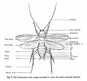
Background imageAnatomy Collection: Main external features of the cockroach. Line engraving

Main external features of the cockroach. Line engraving
COCKROACH. Main external features of the cockroach. Line engraving

Background imageAnatomy Collection: The Zodiac Man - The oldest printed bloodletting chart, showing the astrological signs for

The Zodiac Man - The oldest printed bloodletting chart, showing the astrological signs for bloodletting
BLOODLETTING CHART, 1493. The Zodiac Man - The oldest printed bloodletting chart, showing the astrological signs for bloodletting

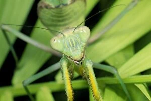
Background imageAnatomy Collection: Praying Mantis

Praying Mantis
SPH-105 Praying Mantis Sphodromantis lineola Steve Hopkin Please note that prints are for personal display purposes only and may not be reproduced in any way

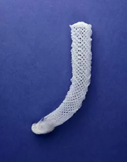
Background imageAnatomy Collection: Euplectella aspergillum, glass sponge

Euplectella aspergillum, glass sponge
Bleached skeleton the a deep-water glass sponge (Euplectella aspergillum). This species can be found attached to rocky parts of the sea floor in the western Pacific near the Philippines

Background imageAnatomy Collection: Parasaurolophus skull

Parasaurolophus skull
A fossil skull belonging to the dinosaur Parasaurolophus on display at the Natural History Museum, London. This large crested dinosaur lived around 70 million years ago during the Upper Cretaceous

Background imageAnatomy Collection: Macrocheira kaempferi, Japanese spider crab

Macrocheira kaempferi, Japanese spider crab
Specimen of the largest crab alive. When fully grown its legs can span amost 4m. It lives at the bottom of the Pacific around Japan

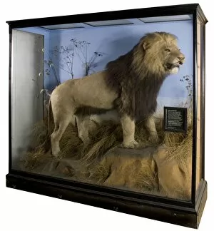
Background imageAnatomy Collection: Panthera leo melanochaitus, cape Lion

Panthera leo melanochaitus, cape Lion
This mounted skin was sent to the Museum in 1954. Shot near South Africas Orange River in about 1830, by Captain Copland-Crawford of the Royal Artillery

Background imageAnatomy Collection: Homo neanderthalensis (Ferrassie 1) cranium cast

Homo neanderthalensis (Ferrassie 1) cranium cast
Cranium cast of an adult male Neandertal Man (Homo neanderthalensis) discovered at La Ferrassie, Dordogne, France, by D Peyrony and L. Captian in 1909. The original specimen dates back 70, 000 years

Background imageAnatomy Collection: Homo sapiens tibia

Homo sapiens tibia
Two human bones from different sides of the Atlantic ocean used to show how syphillis was spread since its appearance in Naples in 1495

Background imageAnatomy Collection: Homo sapiens cranium (Predmosti 3) cast

Homo sapiens cranium (Predmosti 3) cast

Background imageAnatomy Collection: Skull showing bullet hole in right temple

Skull showing bullet hole in right temple
Skull of adult male suicide case. Taken during the archaelogical excavation at Christ Church, Spitalfields, London, 1984-1986

Background imageAnatomy Collection: Homo erectus, Java Man (Sangiran 17) cranium cast

Homo erectus, Java Man (Sangiran 17) cranium cast
Three quarter view of partially reconstructed cranium of Homo erectus Java Man about 700, 000 years old known as Sangiran 17. Discovered by Towikromo in 1969

Background imageAnatomy Collection: Homo erectus crania (Ngandong 1 & Trinil)

Homo erectus crania (Ngandong 1 & Trinil)
The larger cranium belongs to Homo erectus discovered at Ngandong near to the Solo River on the island of Java, Indonesia

Background imageAnatomy Collection: Phobaeticus chani, Chans megastick

Phobaeticus chani, Chans megastick
George Beccaloni holding the Phobaeticus chani specimen. This stick insect from the island of Borneo measures well over a foot in length, and has been identified as the worlds longest insect

Background imageAnatomy Collection: Megalomys luciae, saint lucia giant rice rat (holotype)

Megalomys luciae, saint lucia giant rice rat (holotype). Catalogue number NHM 1853.12.16.2

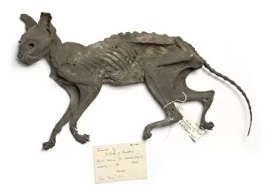
Background imageAnatomy Collection: Naturally mummified Felis catus, cat

Naturally mummified Felis catus, cat
Dried cat found during the demolishing of property on the Duke of Bedfords estate. May 1915. The cat is thought to be approximately 300 years old and had been buried in an air-tight brick-lined cavity

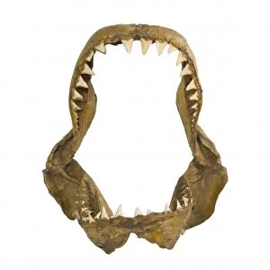
Background imageAnatomy Collection: Carcharodon carcharias, great white shark jaw bones

Carcharodon carcharias, great white shark jaw bones
Specimen of the jaw bones of a great white shark. This species can be found in temperate coastal waters the world over and can grow up to 6m in length

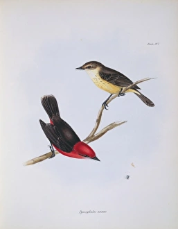
Background imageAnatomy Collection: Pyrocephalus rubinus nanus, vermilion flycatcher

Pyrocephalus rubinus nanus, vermilion flycatcher
Plate 7, hand coloured lithograph by John and Elizabeth Gould from Charles Robert Darwins Zoology of the Voyage of the H.M.S. Beagle, Part 3 Birds, (1838-41). This is not one of Darwins finches

Background imageAnatomy Collection: Verdite head

Verdite head
African head carved in verdite by James Tandi. Verdite is also known as Africas Green Gold and is a member of the Silicates mineral group

Background imageAnatomy Collection: Homo sapiens cranium (Irhoud 1)

Homo sapiens cranium (Irhoud 1)
Lateral and frontal view of a cast of an adult male Homo sapiens skull discovered at Jebel Irhoud (formerly Ighoud), Morocco, by Mohammed Ben Fatmi, Summer 1961

Background imageAnatomy Collection: Columba palumbus, woodpigeon

Columba palumbus, woodpigeon
Plate 23 from William MacGillivrays Watercolour drawings of British Animals (1831-1841)

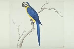
Background imageAnatomy Collection: Ara ararauna, blue-and-yellow macaw

Ara ararauna, blue-and-yellow macaw
Large Series plate 27, a watercolour from the John Reeves Collection of Zoological Drawings from Canton, China

Background imageAnatomy Collection: Limulus polyphemus, horsehoe crab

Limulus polyphemus, horsehoe crab
LS Plate 95 from the John Reeves Collection. John Reeves, a 19th Century Tea Inspector, travelled to Canton, China in order to develop a large collection of Chinese natural history drawings

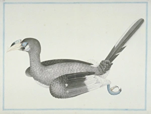
Background imageAnatomy Collection: Anthracoceros albirostris convexus, Oriental pied hornbill

Anthracoceros albirostris convexus, Oriental pied hornbill
Plate 39, painting by Pieter Cornelius de Bevere, from the Loten Collection of coloured drawings of Birds, Mammals, Insects & Plants, (1754-57)

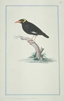
Background imageAnatomy Collection: Gracula religiosa, hill myna

Gracula religiosa, hill myna
Plate 71, painting by Pieter Cornelius de Bevere, from the Loten Collection of coloured drawings of Birds, Mammals, Insects & Plants, (1754-57)

Background imageAnatomy Collection: Plate 90 from the John Reeves Collection

Plate 90 from the John Reeves Collection
John Reeves, a 19th Century Tea Inspector, travelled to Canton, China in order to develop a large collection of Chinese natural history drawings

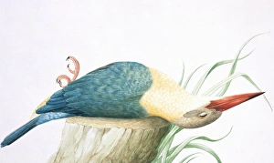
Background imageAnatomy Collection: Pelargopsis capensis, stork-billed kingfisher

Pelargopsis capensis, stork-billed kingfisher
Plate 31, painting by Pieter Cornelius de Bevere, from the Loten Collection of coloured drawings of Birds, Mammals, Insects & Plants, (1754-57)

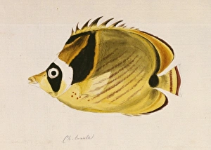
Background imageAnatomy Collection: Chaetodon lunula, raccoon butterflyfish

Chaetodon lunula, raccoon butterflyfish
Ff. 27a Vol 2. Watercolour painting by Sydney Parkinson made during Captain James Cooks first voyage to explore the southern continent 1768-1771

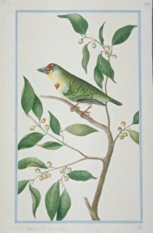
Background imageAnatomy Collection: Megalaima rubricapillus, crimson-fronted barbet

Megalaima rubricapillus, crimson-fronted barbet
Plate 29, painting by Pieter Cornelius de Bevere, from the Loten Collection of coloured drawings of Birds, Mammals, Insects & Plants, (1754-57)


All Professionally Made to Order for Quick Shipping

"Unveiling the Intricacies of Anatomy: From Sensory Homunculus to Motor Homunculus" Step into the fascinating world of anatomy, where every detail tells a story. Explore the depths of human and animal structures as we delve into this captivating subject. Let's begin with Leonardo da Vinci's masterpiece, "Head of a Bear. " Created in 1480 but discovered in 1945, this artwork showcases da Vinci's unparalleled ability to capture anatomical accuracy. The intricate details reveal his keen observation skills and deep understanding of form. Moving on to our furry friends, the greyhound takes center stage. Discovering the anatomy of these majestic creatures unveils their remarkable speed and agility. From their sleek skeleton to their muscular build, it is evident why they are renowned for their racing abilities. But what about us humans? Enter the sensory homunculus – a visual representation that depicts how our brain perceives different body parts based on sensitivity levels. This map reveals intriguing insights into how our senses are distributed throughout our bodies. X-ray images provide another dimension to understanding anatomy. Take a glimpse at normal knees through an X-ray lens – marvel at the complexity hidden beneath our skin while appreciating its delicate balance between strength and flexibility. The backbone is often considered one of nature's most ingenious designs. Our human backbone includes ribs and pelvis, providing stability while allowing movement – truly an architectural marvel worth exploring further. Venturing deeper within ourselves, let us explore the intricacies of the human brain from an inferior view. Witness its complex network of connections responsible for controlling various bodily functions - truly awe-inspiring. As we journey back in time, we encounter hominid crania – remnants that shed light on our evolutionary history. These ancient skulls offer glimpses into early forms such as Australopithecus afarensis (AL 288-1), famously known as Lucy - bridging gaps between past and present.