Fraud Blocker Skip to main content

Animal Cell Collection

"Exploring the Intricate World of Animal Cells

Background imageAnimal Cell Collection: Cell types, artwork

Cell types, artwork
Cell types. Cutaway artwork with a scale bar (upper right, in micrometres) showing the relative sizes of eukaryotic cells (those containing a nucleus) and prokaryotic cells (those lacking a nucleus)

Background imageAnimal Cell Collection: Fibroblast cell, artwork

Fibroblast cell, artwork
Fibroblast cell. Computer artwork of a fibroblast excreting collagen fibres (tropocollagen). Fibroblasts are cells that produce connective tissue such as collagen (tropocollagen)

Background imageAnimal Cell Collection: Animal cell structure

Animal cell structure. Artwork of a section through an animal cell. At lower centre is the nucleus, which contains the cells genetic information in the form of DNA (deoxyribonucleic acid)

Background imageAnimal Cell Collection: Cell, illustration

Cell, illustration
3d illustration of cell division, cell membrane and a splitting red nucleus

Background imageAnimal Cell Collection: Animal cell anatomy, diagram

Animal cell anatomy, diagram
Animal cell anatomy. Diagram showing the internal and external anatomy of an animal cell

Background imageAnimal Cell Collection: Cell of the queen

Cell of the queen

Background imageAnimal Cell Collection: Structure of an animal cell, digital illustration

Structure of an animal cell, digital illustration

Background imageAnimal Cell Collection: Model of animal cell, including cell nucleus, golgi body, lysosomes, centrioles, mitochondria

Model of animal cell, including cell nucleus, golgi body, lysosomes, centrioles, mitochondria, endoplasmic reticulum, ribosomes, cytoplasm, vesicles, thin plasma membrane

Background imageAnimal Cell Collection: Animal cells, artwork F006 / 2225

Animal cells, artwork F006 / 2225
Animal cells, computer artwork

Background imageAnimal Cell Collection: Animal cells, artwork F006 / 2224

Animal cells, artwork F006 / 2224
Animal cells, computer artwork

Background imageAnimal Cell Collection: Animal cells, artwork F006 / 2223

Animal cells, artwork F006 / 2223
Animal cells, computer artwork

Background imageAnimal Cell Collection: Animal cells, artwork F006 / 2222

Animal cells, artwork F006 / 2222
Animal cells, computer artwork

Background imageAnimal Cell Collection: Nucleus and endoplasmic reticulum F006 / 9196

Nucleus and endoplasmic reticulum F006 / 9196
Computer artwork showing part of a human or eukaryotic cell. In the middle the nucleus which has a membrane with nuclear pores. Inside the nucleus is the DNA

Background imageAnimal Cell Collection: Animal cell, artwork F006 / 9019

Animal cell, artwork F006 / 9019
Animal cell, cut-away computer artwork

Background imageAnimal Cell Collection: Nucleus and endoplasmic reticulum F006 / 9201

Nucleus and endoplasmic reticulum F006 / 9201
Computer artwork showing part of a human or eukaryotic cell. In the middle the nucleus which has a membrane with nuclear pores. Inside the nucleus is the DNA

Background imageAnimal Cell Collection: Sodium-potassium ion pump proteins C015 / 9993

Sodium-potassium ion pump proteins C015 / 9993
Sodium-potassium ion pump proteins, molecular model. Sodium-potassium ATPase (adenosine triphosphatase) is an ATP-powered ion pump found in all animal cells

Background imageAnimal Cell Collection: Sodium-potassium ion pump proteins C015 / 9997

Sodium-potassium ion pump proteins C015 / 9997
Sodium-potassium ion pump proteins, molecular model. Sodium-potassium ATPase (adenosine triphosphatase) is an ATP-powered ion pump found in all animal cells

Background imageAnimal Cell Collection: Meiosis, illustration C018 / 0803

Meiosis, illustration C018 / 0803
Meiosis. Illustration of a cell during metaphase (I) of meiosis (gamete formation). During meiosis four daughter nuclei are formed from one parent nucleus after two stages of nuclear division

Background imageAnimal Cell Collection: Animal cell, illustration C018 / 0734

Animal cell, illustration C018 / 0734
Animal cell. Illustration showing the organelles present in an animal cell. At lower centre is the nucleus, which contains the cells genetic information in the form of DNA (deoxyribonucleic acid)

Background imageAnimal Cell Collection: Animal cell organelles, artwork

Animal cell organelles, artwork
Animal cell organelles. Artwork showing the organelles in a eukaryotic cell. This is an animal cell. Structures include the nucleus (centre) which has a membrane with nuclear pores (purple)

Background imageAnimal Cell Collection: Mitochondrion structure, artwork

Mitochondrion structure, artwork
Mitochondrion structure. Artwork showing the internal structure of a mitochondrion. This structure, found in eukaryotic cells, is the site of energy production

Background imageAnimal Cell Collection: Nucleus and endoplasmic reticulum C015 / 6797

Nucleus and endoplasmic reticulum C015 / 6797
Computer artwork showing part of a human or eukaryotic cell. In the middle the nucleus which has a membrane with nuclear pores. Inside the nucleus is the DNA

Background imageAnimal Cell Collection: Nucleus and endoplasmic reticulum C015 / 6783

Nucleus and endoplasmic reticulum C015 / 6783
Computer artwork showing part of a human or eukaryotic cell. In the middle the nucleus which has a membrane with nuclear pores. Inside the nucleus is the DNA

Background imageAnimal Cell Collection: Nucleus and endoplasmic reticulum C015 / 6782

Nucleus and endoplasmic reticulum C015 / 6782
Computer artwork showing part of a human or eukaryotic cell. In the middle the nucleus which has a membrane with nuclear pores. Inside the nucleus is the DNA

Background imageAnimal Cell Collection: Nucleus and endoplasmic reticulum C015 / 6781

Nucleus and endoplasmic reticulum C015 / 6781
Computer artwork showing part of a human or eukaryotic cell. In the middle the nucleus which has a membrane with nuclear pores. Inside the nucleus is the DNA

Background imageAnimal Cell Collection: Nucleus and endoplasmic reticulum C015 / 6772

Nucleus and endoplasmic reticulum C015 / 6772
Computer artwork showing part of a human or eukaryotic cell. In the middle the nucleus which has a membrane with nuclear pores. Inside the nucleus is the DNA

Background imageAnimal Cell Collection: Nucleus and endoplasmic reticulum C015 / 6768

Nucleus and endoplasmic reticulum C015 / 6768
Computer artwork showing part of a human or eukaryotic cell. In the middle the nucleus which has a membrane with nuclear pores. Inside the nucleus is the DNA

Background imageAnimal Cell Collection: Nucleus and endoplasmic reticulum C015 / 6767

Nucleus and endoplasmic reticulum C015 / 6767
Computer artwork showing part of a human or eukaryotic cell. In the middle the nucleus which has a membrane with nuclear pores. Inside the nucleus is the DNA

Background imageAnimal Cell Collection: Sodium-potassium ion pump protein C016 / 2393

Sodium-potassium ion pump protein C016 / 2393
Sodium-potassium ion pump protein, molecular model. Sodium-potassium ATPase (adenosine triphosphatase) is an ATP-powered ion pump found in all animal cells

Background imageAnimal Cell Collection: Sodium-potassium ion pump protein C016 / 2392

Sodium-potassium ion pump protein C016 / 2392
Sodium-potassium ion pump protein, molecular model. Sodium-potassium ATPase (adenosine triphosphatase) is an ATP-powered ion pump found in all animal cells

Background imageAnimal Cell Collection: Animal cell, artwork C013 / 9985

Animal cell, artwork C013 / 9985
Animal cell. Computer artwork showing the cell organelles found inside a typical animal cell. The nucleus (large round) can be seen at centre

Background imageAnimal Cell Collection: Animal cell, artwork C013 / 9984

Animal cell, artwork C013 / 9984
Animal cell. Computer artwork showing the cell organelles found inside a typical animal cell. The nucleus (large round) can be seen at centre

Background imageAnimal Cell Collection: Human cell, artwork C013 / 6522

Human cell, artwork C013 / 6522
Human cell. Illustration of a section through a human cell. At centre is the nucleus (purple), with the nucleolus (dark red) inside it

Background imageAnimal Cell Collection: Cell membrane, artwork C013 / 4988

Cell membrane, artwork C013 / 4988
Cell membrane. Computer artwork of a section through an animal cell showing transmembrane proteins in the cell membrane. The membrane of the cell consists of a dual layer of phospholipids (dark blue)

Background imageAnimal Cell Collection: Cell membrane, artwork C013 / 4986

Cell membrane, artwork C013 / 4986
Cell membrane. Computer artwork of a section through an animal cell showing transmembrane proteins in the cell membrane. The membrane of the cell consists of a dual layer of phospholipids (dark blue)

Background imageAnimal Cell Collection: Golgi apparatus, TEM

Golgi apparatus, TEM
Golgi apparatus. Coloured transmission electron micrograph (TEM) of Golgi apparatus, also known as Golgi bodies, (pink) in a macrophage white blood cell

Background imageAnimal Cell Collection: Inside of an imaginary cell

Inside of an imaginary cell
Inside a cell. Panoramic view from inside an imaginary cell. The densely folded endoplasmic reticulum, a membrane that divides the cell into compartments, is shown in yellow and red

Background imageAnimal Cell Collection: Animal cell structure, computer artwork

Animal cell structure, computer artwork
Animal cell. Computer artwork of a section through an animal cell. At the centre is the nucleus, which contains the cells genetic information in the form of DNA (deoxyribonucleic acid)

Background imageAnimal Cell Collection: Nerve cell anatomy, artwork

Nerve cell anatomy, artwork
Nerve cell anatomy. Computer artwork showing the organelles in a neurone (nerve cell). At centre is nucleus (grey) and nucleolus (black)

Background imageAnimal Cell Collection: Animal cell processes, artwork

Animal cell processes, artwork
Animal cell processes. Cutaway artwork showing the structures inside an animal cell and four different processes that take place inside it or on its membrane (all marked by magnifying glasses)

Background imageAnimal Cell Collection: Animal cell anatomy, artwork

Animal cell anatomy, artwork
Animal cell anatomy. Artwork showing the internal and external anatomy of an animal cell

Background imageAnimal Cell Collection: Animal cell, artwork

Animal cell, artwork
Animal cell. Computer artwork of an expanded animal cell. At centre left is the nucleus (purple sphere), which contains the cells genetic information in the form of DNA (deoxyribonucleic acid)


All Professionally Made to Order for Quick Shipping

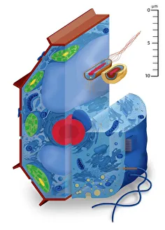
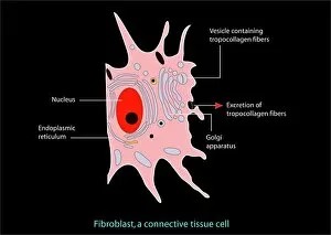
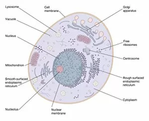
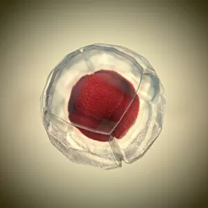
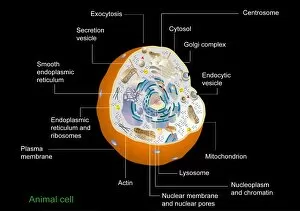
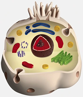
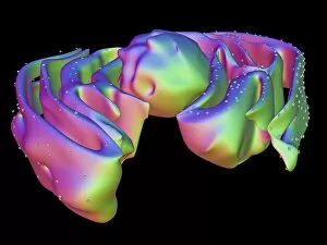
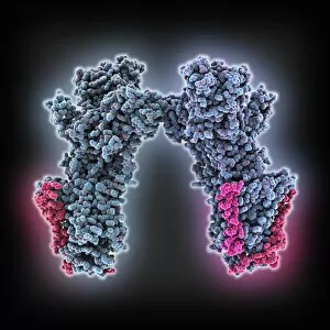
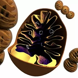
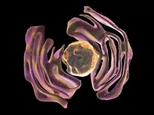
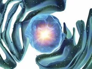
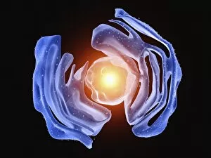
"Exploring the Intricate World of Animal Cells: A Fusion of Art and Science" Delve into the mesmerizing realm of animal cells through captivating artwork that brings these microscopic wonders to life. From fibroblast cells to the majestic queen's cell, each type reveals a unique structure and function. In one stunning illustration, witness the intricate web-like structure of an animal cell, showcasing its nucleus, golgi body, lysosomes, centrioles, mitochondria, endoplasmic reticulum, ribosomes, cytoplasm, vesicles - all enclosed by a thin plasma membrane adorned with microvilli projections at the top. This digital masterpiece unveils the complexity hidden within every single cell. Another artwork captures a newly emerged female worker honey bee pushing out from her brood cell – a testament to nature's remarkable design. The queen's regal cell stands apart in its grandeur and importance within the hive. These artistic renditions not only showcase their aesthetic appeal but also serve as educational tools for understanding animal cells' inner workings. They invite us to marvel at their intricacy while appreciating their vital role in sustaining life. With every stroke of paint or digital brushstroke meticulously applied by talented artists and scientists alike comes an opportunity for us to explore this fascinating world on a new level. These artworks provide visual cues that ignite curiosity and deepen our understanding of these fundamental building blocks of life. Let your imagination soar as you immerse yourself in these breathtaking illustrations depicting various aspects of animal cells' structure and function. Whether it be through detailed models or abstract interpretations, they all contribute to unraveling nature's mysteries right before our eyes. Embark on this artistic journey where science meets creativity; where beauty intertwines with knowledge; where we gain insights into the awe-inspiring complexity residing within each tiny entity known as an animal cell.