
Anthers Collection (page 6)
Anthers: The Intricate Beauty of Flower Stamens Delve into the mesmerizing world of anthers, the vibrant and delicate stamens found in various flowers


All Professionally Made to Order for Quick Shipping
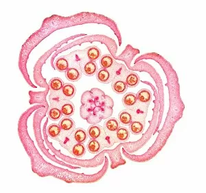
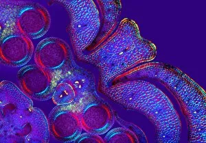
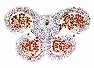
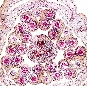
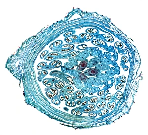
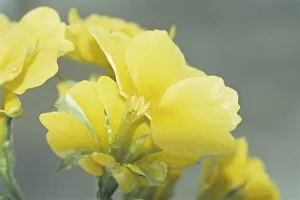
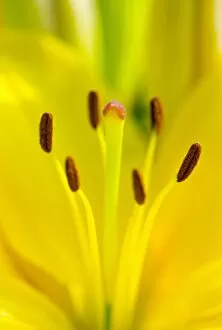
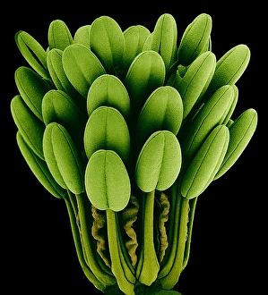
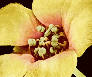
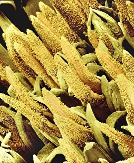
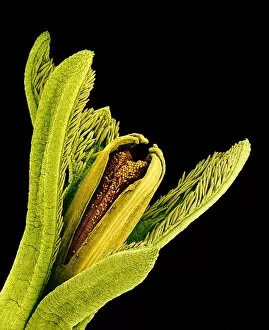
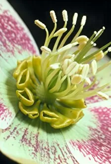
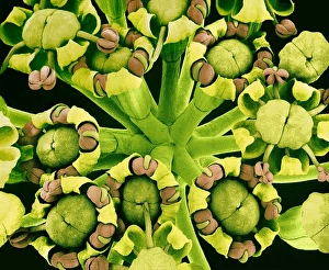
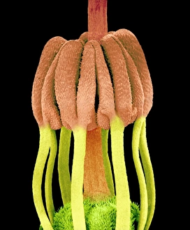
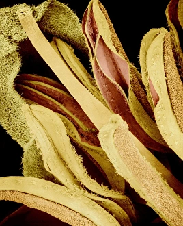
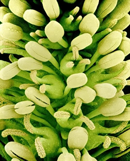
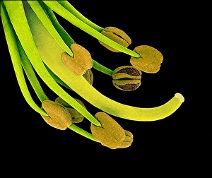
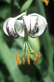
Anthers: The Intricate Beauty of Flower Stamens Delve into the mesmerizing world of anthers, the vibrant and delicate stamens found in various flowers. Through scanning electron microscopy (SEM), we can explore their intricate structures and marvel at their captivating beauty. In the tea flower, anthers take center stage as they release pollen grains during pollination. Under SEM, these tiny structures resemble a colorful tapestry woven with precision and elegance. Moving on to forget-me-not flowers, SEM reveals an enchanting close-up view of their anthers. Each filament seems like a work of art, delicately holding pollen grains that play a vital role in ensuring the plant's reproduction. Traveling to Hong Kong in December brings us face-to-face with the magnificent Hong Kong orchid tree. Its flowers boast stunning anthers that shine under the winter sun, adding a touch of vibrancy to this bustling cityscape. Buttercup flowers also showcase their unique charm through SEM imagery. Their anthers appear as miniature golden orbs nestled within velvety petals - nature's own masterpiece. Columbine flower stamens exhibit fascinating patterns when observed closely under SEM. These intricate designs seem almost otherworldly, inviting us to ponder upon nature's ingenuity. The Amaryllis captures our attention with its majestic blooms adorned by striking anthers. This Hippeastrum species showcases how even reproductive parts can be transformed into objects of sheer beauty. Thale cress offers another glimpse into floral micrography where we witness its minute yet remarkable flower structure through high-resolution imaging techniques. Anther cells become visible like puzzle pieces coming together for life's grand design. Ladys mantle and Euphorbia flowers reveal more secrets when examined using SEM technology. Their reproductive parts astound us with intricacy and diversity - each species showcasing its unique adaptation for successful pollination. Love in the mist displays ethereal beauty through its anthers.