
Arterial Collection
Arterial system, a fascinating network of blood vessels that has captivated scientists and artists alike since the 18th century


All Professionally Made to Order for Quick Shipping
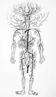
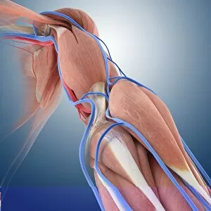
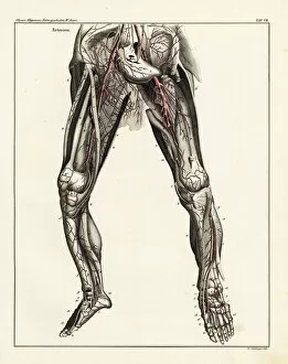
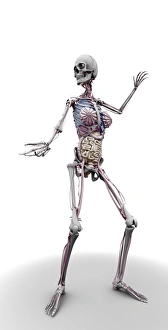
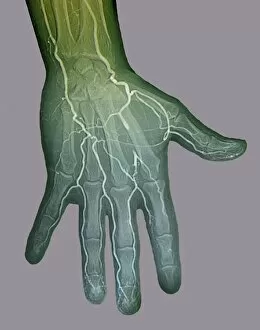
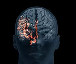
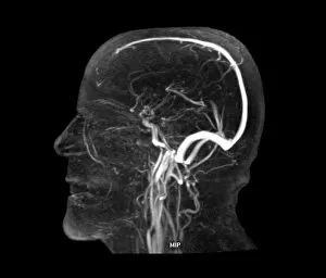
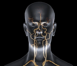
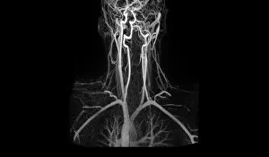
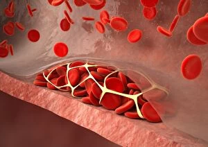
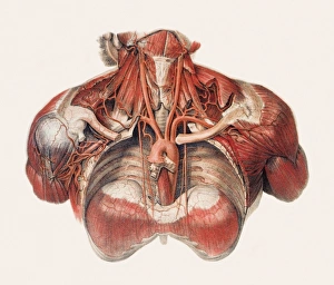
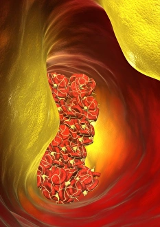
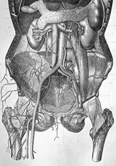
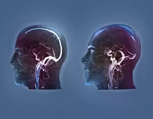
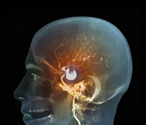
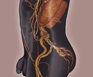
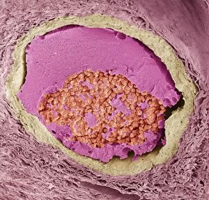
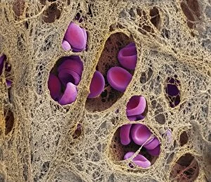
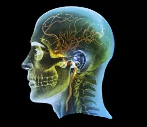
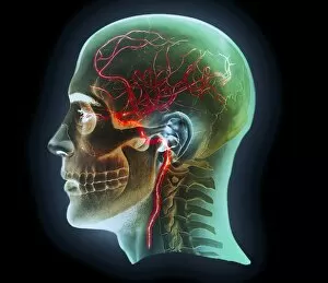
Arterial system, a fascinating network of blood vessels that has captivated scientists and artists alike since the 18th century. From intricate anatomical artwork to modern X-rays, these images provide us with glimpses into the complex world of our circulatory system. In one image, we see the neck and shoulder arteries captured in an X-ray. The delicate branches spread like tree roots, ensuring oxygen-rich blood reaches every corner of our body. Another artwork showcases the arm circulation in stunning detail, highlighting how each vessel intertwines to nourish our muscles and tissues. Moving deeper into the human body, we explore the microscopic wonders within our thyroid gland. Scanning electron microscope (SEM) images reveal capillaries delicately surrounding this vital organ, supplying it with nutrients and hormones essential for proper functioning. The SEM takes us further on this journey as we examine kidney blood vessels intricately woven together like a tapestry. These tiny structures play a crucial role in filtering waste from our bloodstream while maintaining fluid balance within our bodies. Stepping back to admire the bigger picture, we encounter breathtaking artworks depicting the cardiovascular system as a whole. These masterpieces capture both scientific accuracy and artistic beauty as they showcase how arteries branch out from our heart like tributaries flowing through a vast landscape. Delving even deeper into history, we stumble upon heart anatomy illustrations dating back to the 16th century. These ancient depictions remind us of humanity's enduring fascination with understanding this vital organ that keeps us alive. Returning to more recent times, detailed diagrams present an overview systems in different parts of our body - from upper torso to legs - revealing their interconnectedness and importance in sustaining life. And finally, veins and arteries take center stage in an exquisite plate from Recherches Anatomiques Physiologiques focused on head anatomy. This captivating image reminds us that even within such a small area lies an intricate web responsible for delivering oxygen and nutrients to our brain. However, not all is well within these arteries.