
Arteriole Collection
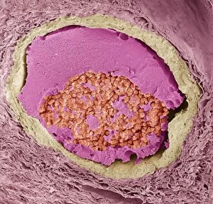
"Exploring the Intricate World of Arterioles: From Thyroid Gland to Kidney Function" Delving into the Thyroid Gland Capillaries


All Professionally Made to Order for Quick Shipping
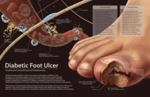
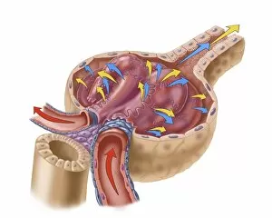
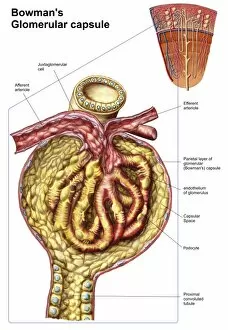
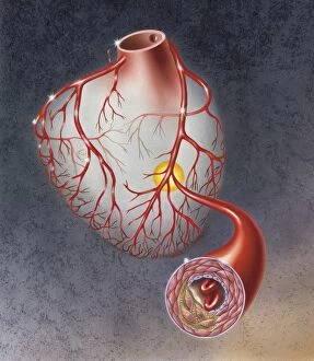
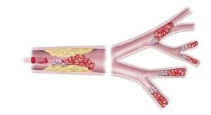
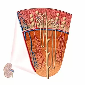
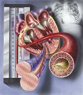
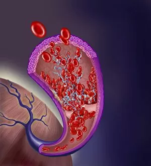
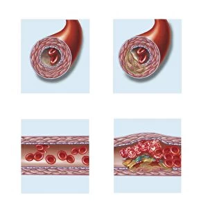
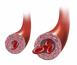
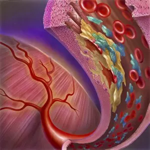
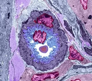
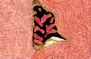
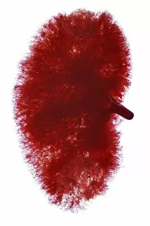
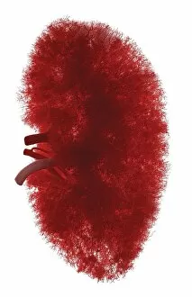
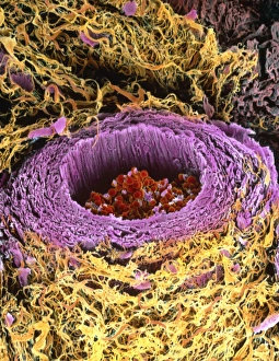
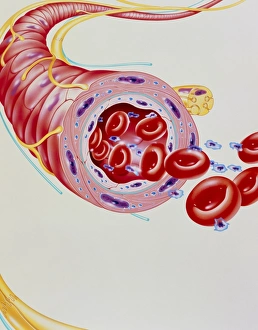
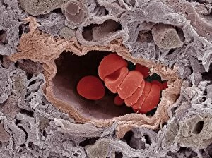
"Exploring the Intricate World of Arterioles: From Thyroid Gland to Kidney Function" Delving into the Thyroid Gland Capillaries: A mesmerizing SEM image reveals the intricate network of arterioles that supply vital nutrients and oxygen to this essential endocrine gland. Unveiling the Complexity of Thyroid Gland Blood Vessels: SEM captures a stunning view, showcasing the branching pattern and microscopic details of arterioles within this crucial gland. Journeying through Kidney Blood Vessels: Witnessing an extraordinary SEM image, we marvel at the labyrinthine pathways formed by arterioles in our kidneys, responsible for maintaining proper filtration and waste removal. Exploring Kidney Glomeruli in Stunning Detail: SEM allows us to appreciate the delicate capillaries within kidney glomeruli, where blood is filtered to produce urine – a true masterpiece of nature's design. Shedding Light on Diabetic Foot Ulcers' Pathophysiology: Investigating histopathology, we unravel how impaired blood flow through damaged arterioles contributes to diabetic foot ulcers' formation and progression. Contrasting Blood Flow Dynamics in Relaxed vs Spasming Arteries: Comparative imagery illustrates how arterial spasm disrupts normal blood flow patterns, emphasizing the importance of healthy arteriole function for overall cardiovascular health. An Artist's Vision of Glomerulus Capillaries: Through an artistic depiction, we gain insight into these tiny vessels' role in filtering waste products from our bloodstream – a testament to their remarkable functionality. Revealing Atherosclerotic Plaque Within Heart Arteries: Examining arteries on a heart model uncovers the detrimental effects of plaque buildup within arterioles – highlighting potential risks associated with poor vascular health. Microvascular Obstruction in Acute Coronary Syndrome Unveiled.