Fraud Blocker Skip to main content

Bacillus Collection (page 3)

"Bacillus: Unveiling the Microscopic World of Bacteria" Delving into the microscopic realm, we encounter a diverse array of bacilli

Background imageBacillus Collection: Pond bacteria C015 / 6371

Pond bacteria C015 / 6371
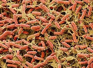
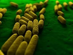
Pond bacteria. Coloured scanning electron micrograph (SEM) of rod-shaped bacteria (bacilli) from a freshwater pond. Magnification: x4000, when printed 10cm wide

Background imageBacillus Collection: Pond bacteria C015 / 6369

Pond bacteria C015 / 6369
Pond bacteria. Coloured scanning electron micrograph (SEM) of rod-shaped bacteria (bacilli) from a freshwater pond. Magnification: x4000, when printed 10cm wide

Background imageBacillus Collection: Propionibacterium acnes C014 / 0634

Propionibacterium acnes C014 / 0634
Propionibacterium acnes bacteria, coloured scanning electron micrograph (SEM). This bacteria is found deep in the hair follicles and pores of the skin

Background imageBacillus Collection: Propionibacterium acnes C014 / 0633

Propionibacterium acnes C014 / 0633
Propionibacterium acnes bacteria, coloured scanning electron micrograph (SEM). This bacteria is found deep in the hair follicles and pores of the skin

Background imageBacillus Collection: Propionibacterium acnes C014 / 0630

Propionibacterium acnes C014 / 0630
Propionibacterium acnes

Background imageBacillus Collection: Actin-based motility, SEM C017 / 8335

Actin-based motility, SEM C017 / 8335
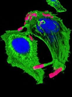
Actin-based motility. Coloured scanning electron micrograph (SEM) of Shigella sp. bacteria (pink) infecting cells. Filaments of the protein actin, which is part of the cells cystoskeleton

Background imageBacillus Collection: Actin-based motility, SEM C017 / 8334

Actin-based motility, SEM C017 / 8334
Actin-based motility. Coloured scanning electron micrograph (SEM) of Shigella sp. bacteria (pink) infecting cells. Filaments of the protein actin, which is part of the cells cystoskeleton

Background imageBacillus Collection: Actin-based motility, light micrograph C017 / 8333

Actin-based motility, light micrograph C017 / 8333
Actin-based motility. 3D model made from optical sections of Shigella sp. bacteria (pink) infecting cells. Filaments of the protein actin, which is part of the cells cystoskeleton

Background imageBacillus Collection: Lactobacillus casei shirota (SEM)

Lactobacillus casei shirota (SEM)
Scanning electron micrograph (SEM) of the bacteria Lactobacillus casei Shirota strain. This particular strain of lactobacillus was isolated by a graduate from the Medical School of Kyoto University

Background imageBacillus Collection: E. coli induced cell death, SEM C016 / 3078

E. coli induced cell death, SEM C016 / 3078
E. coli induced cell death. Coloured scanning electron micrograph (SEM) of a macrophage white blood cell (centre) that is being destroyed by toxins released by Escherichia coli (E)

Background imageBacillus Collection: E. coli induced cell death, SEM C016 / 3077

E. coli induced cell death, SEM C016 / 3077
E. coli induced cell death. Coloured scanning electron micrograph (SEM) of a macrophage white blood cell (centre) that is being destroyed by toxins released by Escherichia coli (E)

Background imageBacillus Collection: EHEC E. coli bacteria, artwork C013 / 4621

EHEC E. coli bacteria, artwork C013 / 4621
EHEC E. coli bacteria. Computer artwork of a enterohaemorrhagic Escherichia coli (EHEC) bacteria in the human gut. E. coli bacteria are a normal part of the intestinal flora in humans

Background imageBacillus Collection: Yoghurt bacteria, SEM

Yoghurt bacteria, SEM
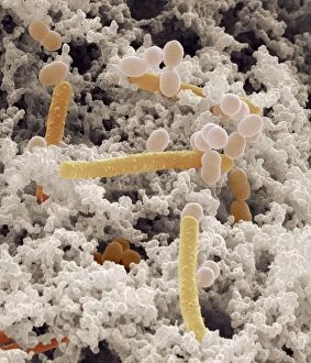
Yoghurt bacteria. Coloured scanning electron micrograph (SEM) of bacteria (coloured) among the milk solids from a yoghurt

Background imageBacillus Collection: Acetobacter bacteria, SEM

Acetobacter bacteria, SEM
Vinegar-forming bacteria. Coloured scanning electron micrograph (SEM) of Acetobacter aceti bacteria, which are used in the production of vinegar

Background imageBacillus Collection: Bacillus sp. bacteria

Bacillus sp. bacteria
Bacteria are the most diverse and ubiquitous soil organisms present on Earth

Background imageBacillus Collection: Stick Insect

Stick Insect
A Stick Insect (Bacillus rossi). Date: 1960s

Background imageBacillus Collection: Food & bacteria in the ileum, SEM

Food & bacteria in the ileum, SEM
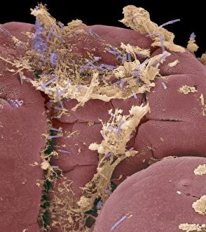
Food and bacteria in the small intestine. Coloured scanning electron micrograph (SEM) of food debris (yellow) and normal intestinal bacteria (purple) on villi (projections, red) in the ileum

Background imageBacillus Collection: Oesophagus epithelium, SEM

Oesophagus epithelium, SEM
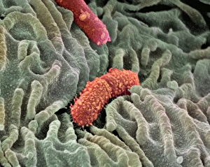
Oesophagus epithelium. Coloured scanning electron micrograph (SEM) of the lining of the human oesophagus or gullet (yellow) and two unknown bacteria (pink & purple)

Background imageBacillus Collection: Oesophagus lining, SEM

Oesophagus lining, SEM
Oesophagus lining. Coloured scanning electron micrograph (SEM) of the lining (epithelium) of the human oesophagus (gullet, blue) and two bacteria (red)

Background imageBacillus Collection: Adenovirus particles and bacterium, TEM

Adenovirus particles and bacterium, TEM
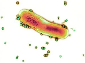
Adenovirus particles and bacterium. Coloured transmission electron micrograph (TEM) of adenovirus particles (green) with a gut bacterium (orange)

Background imageBacillus Collection: Diarrhoea, artwork

Diarrhoea, artwork
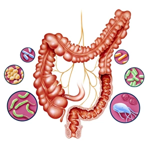
Diarrhoea. Artwork of the human large intestine surrounded by some of the microorganisms that can cause diarrhoea. Diarrhoea is the frequent passing of liquid stools as a symptom of inflammation

Background imageBacillus Collection: Neutrophils and Shigella bacteria, SEM

Neutrophils and Shigella bacteria, SEM
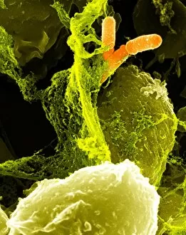
Neutrophils and Shigella bacteria. Coloured scanning electron micrograph (SEM) of neutrophil white blood cells (large, yellow-green) and Shigella bacteria (orange, upper right)

Background imageBacillus Collection: Neutrophil cell and bacteria, SEM

Neutrophil cell and bacteria, SEM
Neutrophil cell and bacteria. Coloured scanning electron micrograph (SEM) of bacteria (rod-shaped) and part of a neutrophil cell (lower left)

Background imageBacillus Collection: Macrophage cell engulfing bacteria, TEM

Macrophage cell engulfing bacteria, TEM
Macrophage cell engulfing bacteria. Coloured transmission electron micrograph (TEM) of bacteria (centre, rod-shaped) inside a macrophage cell

Background imageBacillus Collection: Dividing bacteria, artwork

Dividing bacteria, artwork

Background imageBacillus Collection: Bacillus subtilis bacteria, artwork

Bacillus subtilis bacteria, artwork

Background imageBacillus Collection: Bacteria, conceptual artwork

Bacteria, conceptual artwork

Background imageBacillus Collection: Enterococcus faecalis, bacteria, artwork

Enterococcus faecalis, bacteria, artwork
Enterococcus faecalis. Computer artwork of Enterococcus faecalis bacteria (previously known as Streptococcus faecalis). The bacterium is in the process of dividing through binary fission. E

Background imageBacillus Collection: Bacterial infection, artwork

Bacterial infection, artwork


All Professionally Made to Order for Quick Shipping

"Bacillus: Unveiling the Microscopic World of Bacteria" Delving into the microscopic realm, we encounter a diverse array of bacilli. From the notorious Salmonella bacteria, responsible for foodborne illnesses, to the E. Coli bacterium that can cause severe gastrointestinal infections – these tiny organisms wield significant impact on our health. Through scanning electron microscopy (SEM), we witness astonishing visuals of bacteria found on unexpected surfaces like mobile phones. It serves as a stark reminder to keep our devices clean and germ-free. In H. M. Bateman's intriguing artwork titled "Do you want some?", an artistic representation captures the essence of bacterial transmission through sneezing - highlighting how easily infections can spread in close quarters. Further exploring with transmission electron microscopy (TEM), we observe intricate details of an E. coli bacterium, revealing its complex structure and mechanisms at play. The tuberculosis bacillus emerges as another formidable foe within this microbial world. Artwork depicting both tuberculosis bacteria and bacteriophages showcases their interplay in this ongoing battle against infectious diseases. Taking us back in time, lithographs from 1906 showcase colonies of Haemophilus influenzae and Mycobacterium leprae - providing historical context to our understanding of these pathogens' existence long before modern scientific advancements. Lastly, SEM imagery unveils a captivating view of Salmonella typhimurium bacteria thriving amidst their surroundings; a testament to their resilience and adaptability even under extreme conditions. As we explore the fascinating world of bacilli, it becomes evident that these microorganisms hold immense power over human health. Understanding them better equips us in combating infectious diseases while appreciating their complexity within nature's intricate tapestry.