Fraud Blocker Skip to main content

Bacterial Collection (page 14)

"Bacterial: Unveiling the Microscopic World of Disease and Discovery" Step into a time capsule as we journey through history, exploring the captivating realm of bacteria

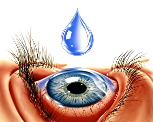
Background imageBacterial Collection: Eye with conjunctivitis

Eye with conjunctivitis
Conjunctivitis. Artwork of an eyedrop being added to an eye suffering from conjunctivitis. This is an inflammation of the conjunctiva

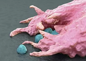
Background imageBacterial Collection: Macrophage engulfing bacteria, artwork

Macrophage engulfing bacteria, artwork
Macrophage engulfing bacteria. Computer artwork of a macrophage white blood cell (pink) engulfing bacteria (blue). This process is called phagocytosis

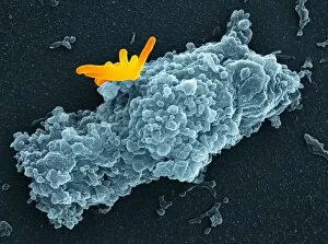
Background imageBacterial Collection: Macrophage engulfing bacteria, SEM

Macrophage engulfing bacteria, SEM
Macrophage engulfing bacteria. Coloured scanning electron micrograph (SEM) of a macrophage white blood cell (brown) engulfing Mycobacterium tuberculosis bacteria (yellow)

Background imageBacterial Collection: Neutrophil cell and bacteria, SEM

Neutrophil cell and bacteria, SEM
Neutrophil cell and bacteria. Coloured scanning electron micrograph (SEM) of bacteria (rod-shaped) and part of a neutrophil cell (lower left)

Background imageBacterial Collection: Macrophage cell engulfing bacteria, TEM

Macrophage cell engulfing bacteria, TEM
Macrophage cell engulfing bacteria. Coloured transmission electron micrograph (TEM) of bacteria (centre, rod-shaped) inside a macrophage cell

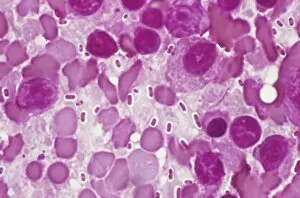
Background imageBacterial Collection: Bacterial pneumonia infection

Bacterial pneumonia infection. Light micrograph of pneumonia bacteria (Klebsiella pneumoniae, rod- shaped, pink) among cells in a sample of pus from a pneumonia patient. K

Background imageBacterial Collection: Kidney infection

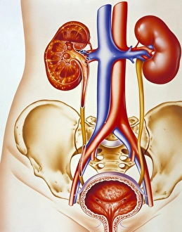
Kidney infection. Artwork of a section through the urinary system showing an infection of a kidney (pyelonephritis, upper left)

Background imageBacterial Collection: Typhoid nodules, light micrograph

Typhoid nodules, light micrograph
Typhoid nodules, coloured light micrograph. Section through a lymph node of a patient with typhoid, showing macrophages (large cells, pink) forming typhoid nodules

Background imageBacterial Collection: Impetigo

Impetigo

Background imageBacterial Collection: Liver tuberculosis, light micrograph

Liver tuberculosis, light micrograph
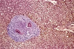
Liver tuberculosis. Coloured light micrograph of a section through the liver of a patient with miliary tuberculosis (TB). A tubercle, a nodular lesion of infected dead tissue, is seen at left

Background imageBacterial Collection: Miliary tuberculosis, light micrograph

Miliary tuberculosis, light micrograph
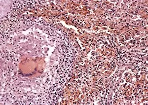
Miliary tuberculosis. Light micrograph of a section through a single miliary tubercle (left) in a patients lung. Tubercles are nodular lesions of infected dead tissue that arise from tuberculosis (TB)

Background imageBacterial Collection: Artwork of meninges & bacteria showing meningitis Artwork of meninges & bacteria showing meningitis

Artwork of meninges & bacteria showing meningitis Artwork of meninges & bacteria showing meningitis
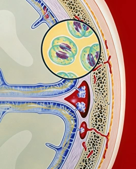
Meningitis. Artwork of a section through the meninges of the brain with an inset picture of Neisseria meningitidis, a cause of meningitis

Background imageBacterial Collection: Stagnant water bacteria, SEM

Stagnant water bacteria, SEM
Stagnant water bacteria. Coloured scanning electron micrograph (SEM) of bacteria attached to a leaf in stagnant water. Numerous proteinaceous filaments extend from the bacteria

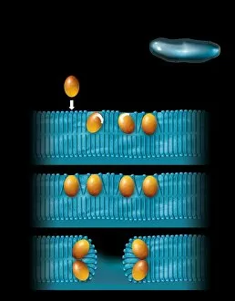
Background imageBacterial Collection: Antibiotic cell membrane effect, artwork

Antibiotic cell membrane effect, artwork
Antibiotic cell membrane effect. Artwork of the natural antibiotic peptide defensin (orange) disrupting the cell membrane of a bacterium (top right)

Background imageBacterial Collection: Astrobiology researcher

Astrobiology researcher Dr Lynn J. Rothschild sampling microbial mats in a lake. These bacteria can survive conditions of high salinity, extreme temperatures and drought

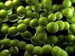
Background imageBacterial Collection: Streptococcus bacteria, artwork

Streptococcus bacteria, artwork

Background imageBacterial Collection: Spirillum bacteria, artwork

Spirillum bacteria, artwork

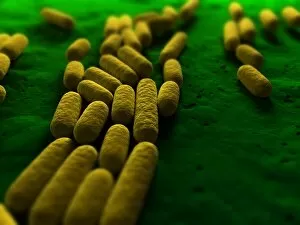
Background imageBacterial Collection: Bacillus subtilis bacteria, artwork

Bacillus subtilis bacteria, artwork

Background imageBacterial Collection: Bacteria, conceptual artwork

Bacteria, conceptual artwork

Background imageBacterial Collection: Enterococcus faecalis, bacteria, artwork

Enterococcus faecalis, bacteria, artwork
Enterococcus faecalis. Computer artwork of Enterococcus faecalis bacteria (previously known as Streptococcus faecalis). The bacterium is in the process of dividing through binary fission. E

Background imageBacterial Collection: Biofactory, conceptual artwork

Biofactory, conceptual artwork
Biofactory, conceptual computer artwork. Cartoon of a factory producing molecules on a conveyor belt. This may represent the use of bacteria to mass-produce molecules for disease prevention

Background imageBacterial Collection: Wound infection, artwork

Wound infection, artwork


All Professionally Made to Order for Quick Shipping

"Bacterial: Unveiling the Microscopic World of Disease and Discovery" Step into a time capsule as we journey through history, exploring the captivating realm of bacteria. From the haunting presence of plague doctors in 17th-century artwork to cutting-edge scientific images captured by scanning electron microscopes (SEM), prepare to be fascinated by these tiny yet formidable organisms. Witness the battle between our immune system and Methicillin-resistant Staphylococcus aureus (MRSA) as a brave neutrophil engulfs it under SEM C018/8596. Marvel at the groundbreaking Calots spinal surgery from the 19th century, which revolutionized medical practices despite lurking bacterial threats. Peering through an X-ray, tuberculosis reveals its sinister grip on human lungs while skin disorders come alive in vivid artwork, reminding us of bacteria's impact on our bodies. E. coli bacteria, magnified under SEM, showcases their intricate structures that belie their harmful potential. Travel back to Soviet Russia in 1967 with a cholera vaccination poster that symbolizes humanity's fight against bacterial epidemics. Observe salmonella bacteria under SEM; their deceptively beautiful appearance belies their ability to cause severe illness. Delve into historical diagrams depicting anthrax cultures and learn about Yersinia pestis - responsible for devastating pandemics throughout history. The tuberculosis bacterium reminds us of its long-standing presence as one of humanity's greatest adversaries. Finally, witness nature's artistic flair with spiral spore chains formed by Streptomyces bacteria – showcasing both beauty and resilience within this microscopic world. Through these glimpses into bacterial realms past and present, we gain insight into our ongoing struggle against infectious diseases. Let this exploration ignite curiosity about microbial lifeforms that shape our existence – forever reminding us how knowledge can empower us in combating these invisible foes.