
Bacteriological Collection
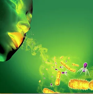
"Bacteriological Wonders: From Infections to Discoveries" Sneezing may seem harmless, but did you know that infections can spread through those tiny droplets


All Professionally Made to Order for Quick Shipping
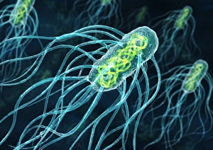
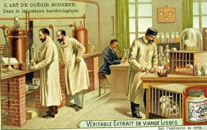
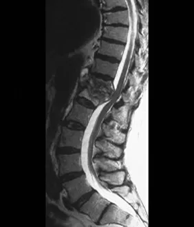
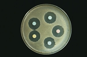
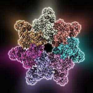
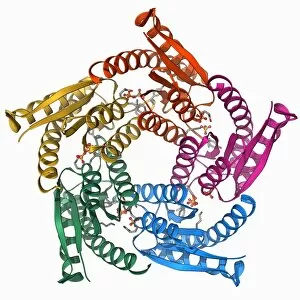
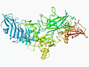
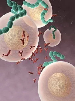
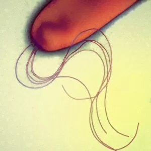
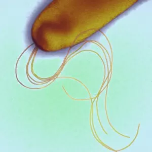
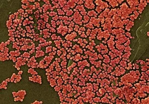
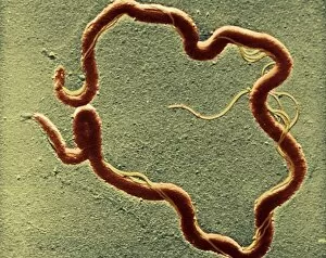
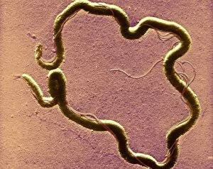
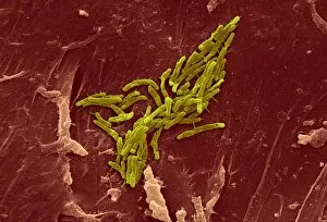
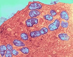
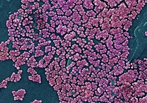
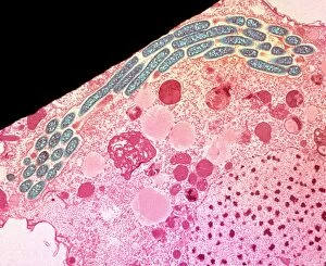
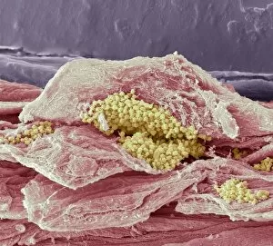
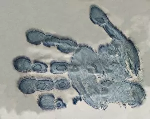
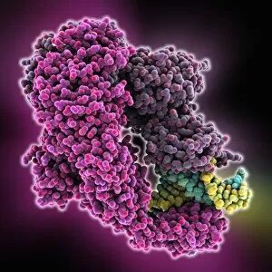
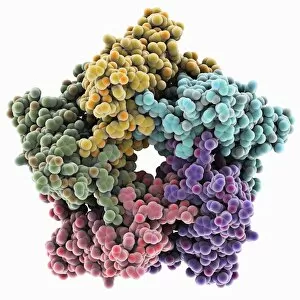
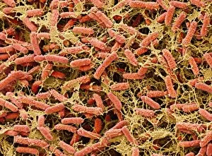
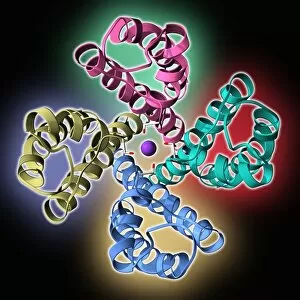
"Bacteriological Wonders: From Infections to Discoveries" Sneezing may seem harmless, but did you know that infections can spread through those tiny droplets? Stay safe and cover your mouth. Take a closer look at artwork C013/5949, showcasing the intricate beauty of Chlamydia trachomatis bacteria under a microscope. Mobile bacteriological laboratories played a crucial role in the medical efforts during World War I, as RAMC officers worked tirelessly in France to combat diseases on the frontlines. Remembering Robert Koch (1843-1910), the German physician who made groundbreaking discoveries in bacteriology and revolutionized our understanding of infectious diseases. Tuberculosis can affect various parts of our body, including the spine. MRI scans like this one help diagnose and treat this challenging condition effectively. Unveiling new possibilities for anthrax treatment, researchers are dedicatedly studying antibiotics to combat this deadly disease. Dive into the world of anthrax research with captivating images like C014/0886 and C014/0865, showcasing protective antigen molecules vital for developing effective vaccines against this lethal infection. The power duo - restriction enzyme and DNA - working together to unlock secrets within genetic material. F006/9315 gives us a glimpse into their fascinating relationship. Lumazine synthase molecule (F006/9291) reveals its critical role in synthesizing essential compounds for bacterial growth – an intriguing aspect of bacteriology worth exploring. Further unraveling anthrax's mysteries is possible with images like F006/9229 and F006/9225 displaying protective antigen molecules that hold immense potential for future treatments against this notorious pathogen. From sneezes spreading infections to remarkable scientific breakthroughs by pioneers like Robert Koch, these glimpses into bacteriology remind us of both the challenges we face and the incredible progress we've made in understanding and combating bacterial diseases.