
Benign Collection
"Exploring the Benign: From Jeremy Bentham to Alphonse III and Beyond" In this captivating journey, we delve into various aspects of the term "benign


All Professionally Made to Order for Quick Shipping
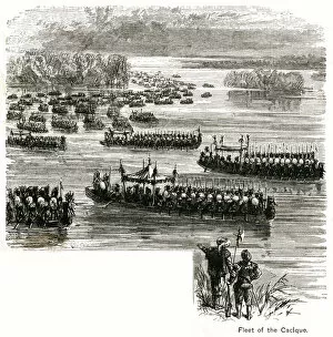
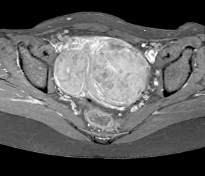
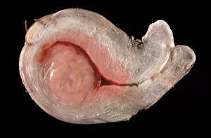
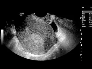
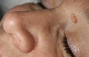
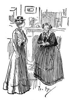
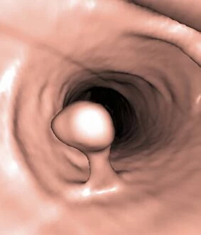
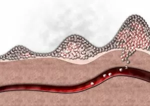
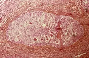
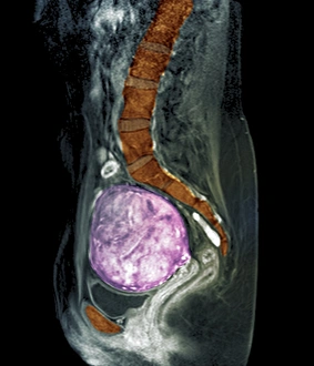
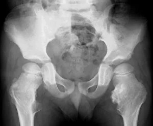
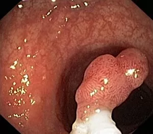
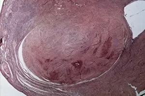
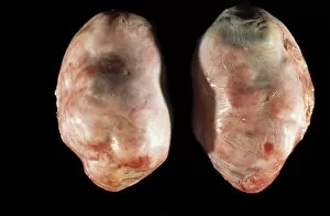
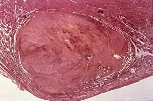
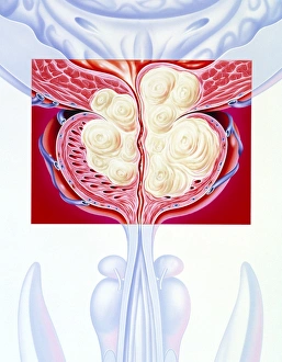
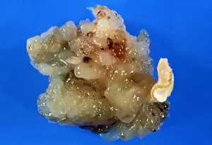
"Exploring the Benign: From Jeremy Bentham to Alphonse III and Beyond" In this captivating journey, we delve into various aspects of the term "benign, " uncovering intriguing connections across different realms. Starting with renowned philosopher Jeremy Bentham, known for his utilitarianism philosophy, we ponder how his ideas shaped our understanding of benevolence in society. Moving on, we encounter Watts' mesmerizing artwork depicting the majestic Fleet of the Cacique Aquixo. Its serene beauty evokes a sense of tranquility and benignity that transports us to another world. Delving into history, we stumble upon Alphonse III The Benign's reign from 1327-1336. As he presided over courts held in Montblanc in 1330, one can't help but wonder about the atmosphere of fairness and justice he brought forth during his rule. Shifting gears to medical insights, an MRI scan (C018/0466) reveals uterine fibroids - a common condition affecting many women worldwide. Despite their presence, these benign growths often pose no serious threat to health. The Akbar Lion Calf Folio found within Shah Jahan Album verso captivates our attention next. This exquisite artwork showcases nature's gentle side as it portrays a lion calf with its mother – a symbol of maternal care and benign protection. Transitioning back to medical terrain, dermal nevi are explored through light micrograph F006/9807. These elevated skin marks remind us that even seemingly harmless features can hold unique stories beneath their surface. Amidst our exploration comes an amusing anecdote when a Lodging House Keeper makes a slight descriptive error. While unintentional, such lighthearted moments remind us that even small mistakes can bring unexpected joy or laughter. Returning to medical discoveries once more, rectal papilloma is examined through a fascinating light micrograph.