
Biofilm Collection
Biofilm is a fascinating phenomenon that can be found in various environments, from dental hygiene to outer space


All Professionally Made to Order for Quick Shipping
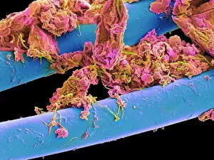
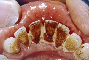
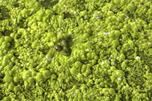
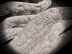
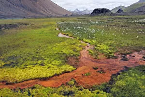
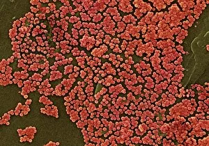
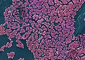
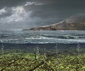
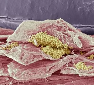
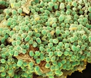
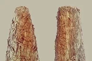
Biofilm is a fascinating phenomenon that can be found in various environments, from dental hygiene to outer space, and is a slimy substance that forms when bacteria adhere to surfaces and create a protective matrix. In the realm of oral health they can be seen when using dental floss. As we glide the floss between our teeth, we may notice a sticky residue being removed - this is dental plaque, which eventually hardens into tartar if not properly addressed. Moving beyond our mouths, it also exists in nature's watery habitats. A close-up view of green slime on a pond at Hawk Island in Georgian Bay, Ontario reveals the intricate structure formed by microorganisms thriving in water. Even frogs are not immune to encountering biofilm. In another close-up shot taken at Georgian Bay, Ontario, we see a frog immersed in slimy green pond water – an environment where biofilms flourish. Interestingly enough, even NASA has delved into studying biofilms for their implications on space exploration. At Kennedy Space Center in Florida, researchers like Jason Fischer and Brint Bauer have been investigating how these films develop and behave under unique conditions. Using scanning electron microscopy (SEM), scientists examine samples collected from water tanks filled with green dye as part of their studies at Kennedy Space Center's Air and Water Revitalization lab. These tanks have spent five years floating through space. Carolina Franco and Christina Khodadad are among the experts who conduct biological studies on these two tanks of water gathered from outer space missions. Their research aims to understand how microorganisms adapt within enclosed systems during long-duration space travel. The study of biofilms provides valuable insights into microbial life and its ability to survive extreme conditions both here on Earth and beyond our planet's atmosphere.