Fraud Blocker Skip to main content

Biomolecule Collection (page 14)

"Biomolecules: Unveiling the Intricate World of Life's Building Blocks" Peering through the lens of scientific discovery

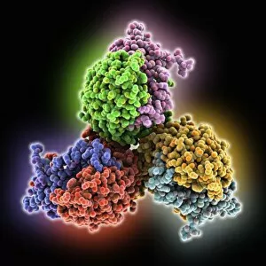
Background imageBiomolecule Collection: Follicle-stimulating hormone complex C016 / 2794

Follicle-stimulating hormone complex C016 / 2794
Follicle-stimulating hormone (FSH) complex with receptor, molecular model. FSH helps to regulate human sexual development and reproductive processes. In females, it acts on follicles in the ovaries

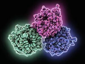
Background imageBiomolecule Collection: RACK1 protein C016 / 2792

RACK1 protein C016 / 2792
RACK1 protein, molecular model. RACK1 stands for Receptor for Activated C Kinase 1. This protein is also known as guanine nucleotide-binding protein subunit beta-2-like 1 (GNB2L1)

Background imageBiomolecule Collection: Follicle-stimulating hormone complex C016 / 2793

Follicle-stimulating hormone complex C016 / 2793
Follicle-stimulating hormone (FSH) complex with receptor, molecular model. FSH helps to regulate human sexual development and reproductive processes. In females, it acts on follicles in the ovaries

Background imageBiomolecule Collection: RACK1 protein C016 / 2791

RACK1 protein C016 / 2791
RACK1 protein, molecular model. RACK1 stands for Receptor for Activated C Kinase 1. This protein is also known as guanine nucleotide-binding protein subunit beta-2-like 1 (GNB2L1)

Background imageBiomolecule Collection: Human tumour protein p63 C016 / 2681

Human tumour protein p63 C016 / 2681
Human tumour protein p63. Molecular model of the tetramerization domain of the human tumour protein p63, encoded by the TP63 gene

Background imageBiomolecule Collection: GMP synthetase enzyme C016 / 2679

GMP synthetase enzyme C016 / 2679
GMP synthetase enzyme, molecular model. This enzyme, guanine monophosphate synthetase, catalyses the reaction that converts xanthosine monophosphate to guanosine monophosphate

Background imageBiomolecule Collection: Human tumour protein p63 C016 / 2680

Human tumour protein p63 C016 / 2680
Human tumour protein p63. Molecular model of the tetramerization domain of the human tumour protein p63, encoded by the TP63 gene

Background imageBiomolecule Collection: GMP synthetase enzyme C016 / 2678

GMP synthetase enzyme C016 / 2678
GMP synthetase enzyme, molecular model. This enzyme, guanine monophosphate synthetase, catalyses the reaction that converts xanthosine monophosphate to guanosine monophosphate

Background imageBiomolecule Collection: RNA-silencing protein with RNA C016 / 2558

RNA-silencing protein with RNA C016 / 2558
RNA-silencing protein with RNA. Molecular model of RNA silencing taking place by human piwi-like protein (purple) acting on short loops (green) of RNA (ribonucleic acid)

Background imageBiomolecule Collection: RNA-silencing protein with RNA C016 / 2557

RNA-silencing protein with RNA C016 / 2557
RNA-silencing protein with RNA. Molecular model of RNA silencing taking place by human piwi-like protein (green) acting on short loops (red) of RNA (ribonucleic acid)

Background imageBiomolecule Collection: Plant anion channel protein homologue C016 / 2555

Plant anion channel protein homologue C016 / 2555
Plant anion channel protein homologue, molecular model. Obtained from the bacterium Haemophilus influenzae, this anion channel protein (TehA) is being studied due to its common structure (homologue)

Background imageBiomolecule Collection: Plant anion channel protein homologue C016 / 2556

Plant anion channel protein homologue C016 / 2556
Plant anion channel protein homologue, molecular model. Obtained from the bacterium Haemophilus influenzae, this anion channel protein (TehA) is being studied due to its common structure (homologue)

Background imageBiomolecule Collection: Sodium-potassium ion pump protein C016 / 2393

Sodium-potassium ion pump protein C016 / 2393
Sodium-potassium ion pump protein, molecular model. Sodium-potassium ATPase (adenosine triphosphatase) is an ATP-powered ion pump found in all animal cells

Background imageBiomolecule Collection: Sodium-potassium ion pump protein C016 / 2392

Sodium-potassium ion pump protein C016 / 2392
Sodium-potassium ion pump protein, molecular model. Sodium-potassium ATPase (adenosine triphosphatase) is an ATP-powered ion pump found in all animal cells

Background imageBiomolecule Collection: Dengue virus protein and antibody C016 / 2375

Dengue virus protein and antibody C016 / 2375
Dengue virus protein and antibody. Molecular model of a dengue virus 2 envelope protein (yellow) complexed with antibody Mab 4E11 (purple)

Background imageBiomolecule Collection: Helicase transcriptional silencer protein C016 / 2376

Helicase transcriptional silencer protein C016 / 2376
Helicase transcriptional silencer protein. Molecular model of the helicase protein MOM1 acting as a transcriptional silencer

Background imageBiomolecule Collection: Dengue virus protein and antibody C016 / 2374

Dengue virus protein and antibody C016 / 2374
Dengue virus protein and antibody. Molecular model of a dengue virus 2 envelope protein (red) complexed with antibody Mab 4E11 (blue)

Background imageBiomolecule Collection: Transthyretin blood protein C016 / 2327

Transthyretin blood protein C016 / 2327
Transthyretin blood protein. Molecular model of the thyroid hormone binding protein transthyretin, also known as prealbumin

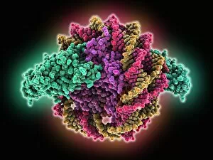
Background imageBiomolecule Collection: Sir3 gene silencer acting on DNA C016 / 2325

Sir3 gene silencer acting on DNA C016 / 2325
Sir3 gene silencer acting on DNA, molecular model. Sir3 (bright green) is acting on a circular strand of DNA (deoxyribonucleic acid, red and yellow)

Background imageBiomolecule Collection: Sir3 gene silencer acting on DNA C016 / 2324

Sir3 gene silencer acting on DNA C016 / 2324
Sir3 gene silencer acting on DNA, molecular model. Sir3 (light blue) is acting on a circular strand of DNA (deoxyribonucleic acid, pink)

Background imageBiomolecule Collection: Transthyretin blood protein C016 / 2326

Transthyretin blood protein C016 / 2326
Transthyretin blood protein. Molecular model of the thyroid hormone binding protein transthyretin, also known as prealbumin

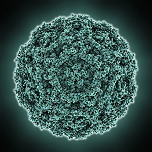
Background imageBiomolecule Collection: B19 parvovirus capsid protein

B19 parvovirus capsid protein, molecular model. This protein forms part of the capsid (protein shell) of parvovirus B19, one of only a few parvoviruses that infect humans

Background imageBiomolecule Collection: Heterotrimeric G protein complex C013 / 7186

Heterotrimeric G protein complex C013 / 7186
Heterotrimeric G protein complex, molecular model showing secondary structure. Also called the large G proteins, these activate enzymes and metabolic pathways

Background imageBiomolecule Collection: Vitamin B12 transport protein C013 / 7184

Vitamin B12 transport protein C013 / 7184
Vitamin B12 transport protein, molecular model showing secondary structure. This receptor protein, known as BTUB, is from the Escherichia coli bacterium

Background imageBiomolecule Collection: FKBP52 immunoregulation enzyme C013 / 7182

FKBP52 immunoregulation enzyme C013 / 7182
FKBP52 immunoregulation enzyme, molecular model showing secondary structure. This protein, encoded by the human FKBP4 gene, is a type of enzyme called a prolyl isomerase

Background imageBiomolecule Collection: Escherichia coli heat-labile enterotoxin C013 / 7180

Escherichia coli heat-labile enterotoxin C013 / 7180
Escherichia coli heat-labile enterotoxin, molecular model showing secondary structure. This is one of several proteins produced by pathogenic E. coli bacteria in the intestines

Background imageBiomolecule Collection: Bacteriophage MS2 capsid protein C013 / 7181

Bacteriophage MS2 capsid protein C013 / 7181
Bacteriophage MS2 capsid protein, molecular model showing secondary structure. This protein forms part of the capsid (protein shell) of MS2

Background imageBiomolecule Collection: Nitrogen-fixing molybdenum iron enzyme C013 / 7176

Nitrogen-fixing molybdenum iron enzyme C013 / 7176
Nitrogen-fixing molybdenum iron enzyme, molecular model showing secondary structure. This protein is a nitrogen fixation enzyme (nitrogenase)

Background imageBiomolecule Collection: Human prion precursor protein C013 / 7178

Human prion precursor protein C013 / 7178
Human prion precursor protein, molecular model showing secondary structure. The human prion protein (hPrP) is a prion precursor

Background imageBiomolecule Collection: Fatty acid dehydrogenase enzyme C013 / 7175

Fatty acid dehydrogenase enzyme C013 / 7175
Fatty acid dehydrogenase enzyme, molecular model showing secondary structure. This is short chain 3-hydroxyacyl CoA dehydrogenase (SCHAD)

Background imageBiomolecule Collection: Clostridium botulinum toxin fragment C013 / 7173

Clostridium botulinum toxin fragment C013 / 7173
Clostridium botulinum toxin fragment, molecular model showing secondary structure. This binding domain fragment (BBHc), a hydrolase, is from botulinum neurotoxin type B

Background imageBiomolecule Collection: HIV-1 reverse transcriptase enzyme C013 / 7172

HIV-1 reverse transcriptase enzyme C013 / 7172
HIV-1 reverse transcriptase enzyme, molecular model showing secondary structure. This protein is an enzyme that mediates the copying of genetic information

Background imageBiomolecule Collection: Molecule in space

Molecule in space, conceptual computer artwork. The theory of panspermia states that the molecules that form the building blocks of life are found throughout the universe

Background imageBiomolecule Collection: Cryptochrome and rhodopsin pigments

Cryptochrome and rhodopsin pigments. Rhodopsin is a pigment found in the rod photoreceptor cells in the retina of the human eye, where it absorbs light falling on the retina

Background imageBiomolecule Collection: Heme group in haemoglobin, diagram

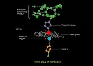
Heme group in haemoglobin, diagram
Heme group in haemoglobin. Diagram showing the heme group (centre) in the haemoglobin molecule (partly shown), the chemical unit that is responsible for the transport of oxygen in the blood

Background imageBiomolecule Collection: Glycogen units, molecular model

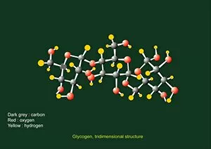
Glycogen units, molecular model. Glycogen is made from many glucose molecules linked by one of two types of glycosidic bonds

Background imageBiomolecule Collection: Glycogen phosphorylase, molecular model

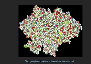
Glycogen phosphorylase, molecular model. This is an enzyme involved in breaking down glycogen, the energy storage molecule involved in animal metabolism

Background imageBiomolecule Collection: Earths cometary bombardment, artwork

Earths cometary bombardment, artwork
Earths cometary bombardment. Artwork of Earth being bombarded with cometary fragments. On a populated Earth, such a bombardment would be catastrophic, but in the early history of the solar system


All Professionally Made to Order for Quick Shipping

"Biomolecules: Unveiling the Intricate World of Life's Building Blocks" Peering through the lens of scientific discovery, we witness a microscopic view of the human respiratory syncytial virus, unraveling its intricate structure and shedding light on its mechanisms. Zinc fingers delicately embrace a DNA strand, forming an elegant dance between proteins and genetic material. This interaction holds the key to unlocking the mysteries encoded within our genes. The SARS coronavirus protein stands as a formidable foe in our battle against infectious diseases. Understanding its structure is crucial for developing effective treatments and vaccines. Illustrated with precision, a protein takes center stage, showcasing nature's artistic design. Its complex folds and twists hold immense power in shaping life's diverse forms and functions. TFAM transcription factor gracefully binds to DNA C015/7059, orchestrating gene expression like a conductor leading an orchestra. This molecular symphony plays a vital role in maintaining cellular harmony. Stylized rabies virus particles remind us of nature's ability to create both beauty and danger simultaneously. The intricacies hidden within these tiny entities have captivated scientists for centuries. A glimpse into the microscopic world reveals yellow fever virus particles resembling vibrant orbs dancing amidst darkness—a reminder that even invisible threats can wield significant impact on human health. Conceptual imagery portrays the enigmatic rabies virus—an embodiment of fear lurking in shadows yet inspiring relentless pursuit towards understanding this deadly pathogen. MyoD muscle protein-DNA complex showcases how molecules choreograph muscle development—binding together like partners engaged in an exquisite ballet that shapes our physical strength. MscL ion channel protein structure presents itself as nature's gateway—allowing ions to traverse cell membranes with precise control over vital processes essential for life itself. Adenovirus hexon protein emerges as an architectural masterpiece—a structural cornerstone dictating viral entry into host cells while captivating researchers seeking innovative antiviral strategies.