
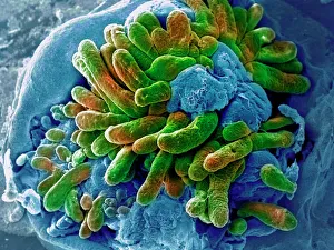
Biopsy Collection
"Exploring the Depths: Unveiling the Secrets of Biopsy" Delving into the Unknown: Unraveling the Mysteries of Small Intestine with SEM A Needle's Journey


All Professionally Made to Order for Quick Shipping
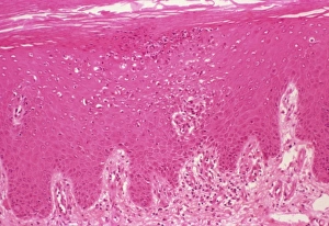
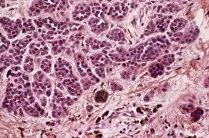
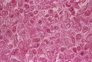
"Exploring the Depths: Unveiling the Secrets of Biopsy" Delving into the Unknown: Unraveling the Mysteries of Small Intestine with SEM A Needle's Journey: Black and White Illustration in Quadriceps Muscle Peering Inside: Cross Section Biomedical Illustration Reveals Prostate Gland Needle Biopsy Procedure Navigating Precision: Endometrial Biopsy Unveiled through Novak Curette, a Cross Sectional Perspective Safeguarding Women's Health: Cervical Conization Biopsy Sheds Light on Cervical Intraepithelial Neoplasia Beneath the Surface: Exploring Skin Tissues with a Cross Sectional Site of Skin Biopsy Microscopic Insights: Examining Tissue Samples - The Fascinating World Slides F007 / 0316 Battling CMV Lung Infection - Understanding its Impact through a Microscopic Lens Shedding Light on Benign Testicle Tumors - Illuminating Discoveries from Light Micrograph Analysis Bone Deep Exploration - Analyzing Bone Biopsy Samples for Deeper Medical Insight Visualizing Beyond Limits.