Fraud Blocker Skip to main content

Blastomere Collection

A blastomere is a crucial component in the early stages of embryonic development

Background imageBlastomere Collection: Cluster of blastomeres forming a developing morula

Cluster of blastomeres forming a developing morula (early stage of embryonic development)

Background imageBlastomere Collection: LM of four-cell embryo

LM of four-cell embryo
Four-cell embryo. Light micrograph of the blasto- meres of a four-cell human embryo, two days after fertilisation. The blastomeres are the rounded cells

Background imageBlastomere Collection: Coloured LM of a human embryo at two-cell stage

Coloured LM of a human embryo at two-cell stage

Background imageBlastomere Collection: LM of human embryo at four-cell stage

LM of human embryo at four-cell stage
Light micrograph (Kohler illumination) of a primitive human embryo, composed of four cells following the initial mitotic divisions which ultimately transform a single cell organism into one composed

Background imageBlastomere Collection: LM of human embryo at two-cell stage

LM of human embryo at two-cell stage
Light micrograph of a human zygote (the primitive embryo), composed of two cells following its first mitotic division - the initial transformation from a single cell organism to one composed of

Background imageBlastomere Collection: LM of blastomeres of a two-cell embryo

LM of blastomeres of a two-cell embryo
Two-cell embryo. Light micrograph of the blasto- meres of a 2-cell human embryo, two days after fertilisation. The blastomeres are the rounded cells formed by the first division of the fertilized

Background imageBlastomere Collection: LM of human embryo at eight cell stage

LM of human embryo at eight cell stage
Light micrograph of a primitive human embryo, composed of eight cells following the initial mitotic divisions which ultimately transform a single cell organism into one composed of millions of cells

Background imageBlastomere Collection: Coloured SEM of human embryo at 8-cell stage

Coloured SEM of human embryo at 8-cell stage
Eight-cell embryo. Coloured scanning electron micrograph (SEM) of a human embryo at the eight cell stage, three days after fertilisation

Background imageBlastomere Collection: Vertebrate embryonic development, artwork

Vertebrate embryonic development, artwork
Vertebrate embryonic development. Historical artwork showing the development of an embryo from cleavage (top left) to gastrulation (bottom right)


All Professionally Made to Order for Quick Shipping

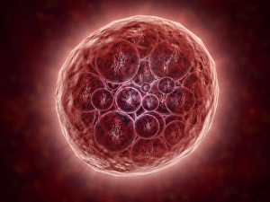
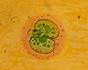
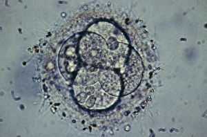
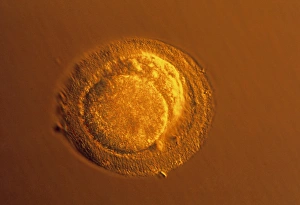
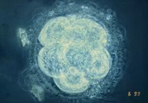
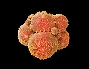
A blastomere is a crucial component in the early stages of embryonic development. It refers to a cluster of cells that form during the process, creating what is known as a developing morula. In this captivating image, we can observe a beautifully captured light microscope view of a four-cell embryo. The vibrant colors and intricate details showcase the complexity and wonder of life at its earliest stages. Moving closer into the world of human embryos, we are presented with another colored light microscope image depicting an exquisite two-cell stage embryo. The delicate structures within each cell are clearly visible, giving us insight into the remarkable intricacies involved in human development. Zooming even further into this microscopic realm, we encounter an astonishing close-up view of blastomeres within a two-cell embryo. This highly magnified light microscope image allows us to appreciate the individuality and uniqueness present within each tiny cell. As time progresses, so does embryonic growth. Here we witness yet another fascinating glimpse into human development at its four-cell stage through an impressive light microscope photograph. Each cell seems to be bursting with potential and promise for future growth and differentiation. Advancing further along this incredible journey, we come across an awe-inspiring eight-cell stage human embryo under examination using both light microscopy and scanning electron microscopy techniques. The resulting colored SEM image showcases not only the structural beauty but also provides valuable insights into cellular organization during this critical phase. Stepping back from specific images now, let's explore some artwork representing vertebrate embryonic development throughout history. These artistic renderings capture various stages of growth in mesmerizing detail while reminding us how far our understanding has evolved over time. These captivating images and artworks offer glimpses into the extraordinary world of blastomeres and their role in shaping life itself during early embryonic development. They serve as reminders that even on such minuscule scales lies immense beauty waiting to be discovered by those who dare to look closely.