
Bleb Collection
"Bleb: Unveiling the Intricate World of Cancer Cells" In the realm of apoptosis and cancer research, blebs take center stage


All Professionally Made to Order for Quick Shipping
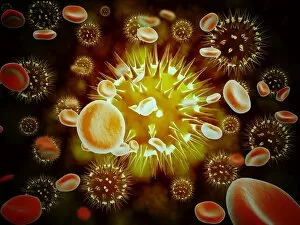
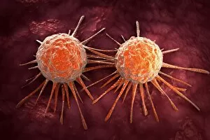
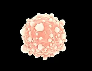
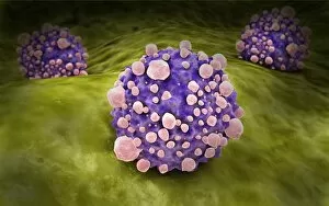
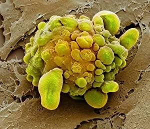
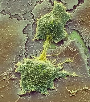
"Bleb: Unveiling the Intricate World of Cancer Cells" In the realm of apoptosis and cancer research, blebs take center stage. These protrusions on cell membranes provide a fascinating glimpse into the intricate mechanisms underlying cellular death and survival. Through scanning electron microscopy (SEM), we delve into a captivating scene where cancer cells intertwine with red blood cells, symbolizing their invasive nature. This mesmerizing image serves as a stark reminder of the relentless battle waged within our bodies. A microscopic view reveals pancreatic cancer cells in all their malignant glory. The intricate patterns they form highlight their aggressive proliferation, emphasizing the urgent need for effective treatments against this devastating disease. Conceptual images depicting cancer viruses further emphasize the grave threat posed by these stealthy invaders. Their menacing presence reminds us that understanding viral interactions is crucial to developing targeted therapies that can halt their progression. Amidst this chaos, we witness another microscopic view showcasing macrophages – our body's frontline defenders against cancerous growths. These immune warriors tirelessly patrol, engulfing and neutralizing aberrant cells in an effort to maintain harmony within our systems. The recurring theme of pancreatic cancer cells underscores its significance as one of the most challenging forms of malignancy faced today. It urges scientists and medical professionals alike to intensify efforts towards early detection methods and innovative treatment strategies. As we navigate through these awe-inspiring glimpses into cellular landscapes, it becomes evident that unraveling the mysteries surrounding blebs holds immense potential for advancing our understanding of apoptosis regulation and combating cancers effectively. "Bleb" encapsulates both beauty and menace within its confines – from captivating SEM images capturing red blood cell flow amidst invading cancer cells to conceptual representations illustrating virus-induced havoc. Through exploring microscopic views featuring macrophages valiantly defending against malignant intruders or clusters of pancreatic cancer cells proliferating relentlessly; we gain valuable insights into this complex world while fueling hope for breakthroughs in cancer research.