Fraud Blocker Skip to main content

Bloodstream Collection

"Journey of Life: Exploring the Intricate Pathways of the Bloodstream" Step back in time to 1861

Background imageBloodstream Collection: Human circulatory system, hand-coloured engraving, published in 1861

Human circulatory system, hand-coloured engraving, published in 1861
The human circulatory system. Hand-coloured engraving, published in 1861

Background imageBloodstream Collection: Diagram of human lungs showing blood supply

Diagram of human lungs showing blood supply

Background imageBloodstream Collection: Diagram showing bloodstream inside a vein, red, white blood cells, platelet

Diagram showing bloodstream inside a vein, red, white blood cells, platelet

Background imageBloodstream Collection: Direct sphygmograph, 1882 (engraving)

Direct sphygmograph, 1882 (engraving)
8766934 Direct sphygmograph, 1882 (engraving); Private Collection; (add.info.: Direct sphygmograph. Dr. Jaccoud-V 33 1882); Photo ©Jaime Abecasis.

Background imageBloodstream Collection: Superficial veins of the skull and face. View side 1905 (engraving)

Superficial veins of the skull and face. View side 1905 (engraving)
7141220 Superficial veins of the skull and face. View side 1905 (engraving); Private Collection; (add.info.: Superficial veins of the skull and face)

Background imageBloodstream Collection: Transmission sphymograph, new model, 1882 (engraving)

Transmission sphymograph, new model, 1882 (engraving)
8766937 Transmission sphymograph, new model, 1882 (engraving); Private Collection; (add.info.: Transmission sphymograph, new model by Marey Dr. Jaccoud V 33 1882); Photo ©Jaime Abecasis

Background imageBloodstream Collection: Yellow ground, red heart

Yellow ground, red heart
E. de Juan

Background imageBloodstream Collection: Illustration from Fasciculus medicinae Venice, 15th century (woodcut)

Illustration from Fasciculus medicinae Venice, 15th century (woodcut)
884990 Illustration from Fasciculus medicinae Venice, 15th century (woodcut) by Italian School, (15th century); Biblioteca Civica, Padua

Background imageBloodstream Collection: Anatomy of the human bloodstream, lithograph, published in 1874

Anatomy of the human bloodstream, lithograph, published in 1874
Anatomy of the human bloodstream. Lithograph, published in 1874

Background imageBloodstream Collection: Anatomy of the human fetus, lithograph, published in 1875

Anatomy of the human fetus, lithograph, published in 1875
Anatomy of the human fetus. Woodcut engraving, published in 1875

Background imageBloodstream Collection: Histopathology and pathophysiology of diabetic food ulcers

Histopathology and pathophysiology of diabetic food ulcers

Background imageBloodstream Collection: Human male anatomy

Human male anatomy
Antique illustration of a Human male anatomy

Background imageBloodstream Collection: Blood flow diagram

Blood flow diagram

Background imageBloodstream Collection: Pair of human lungs

Pair of human lungs

Background imageBloodstream Collection: Fight-or-flight response, artwork

Fight-or-flight response, artwork
Fight-or-flight response. Artwork showing the bodys fight-or-flight response to a threat. Visual perception (yellow arrow) can trigger fear memories controlled by the brains amygdala (yellow)

Background imageBloodstream Collection: Coagulation factor complex molecule C014 / 0139

Coagulation factor complex molecule C014 / 0139
Coagulation factor complex molecule. Molecular model showing the interaction between coagulation factor VIII (FVIII, pink, blue and yellow) and factor IXa (FIXa, cream and grey)

Background imageBloodstream Collection: Illustration showing excess Cholesterol in bloodstream over-saturating body cells

Illustration showing excess Cholesterol in bloodstream over-saturating body cells

Background imageBloodstream Collection: Heart with arteries showing cholesterol in one and plaque in the other

Heart with arteries showing cholesterol in one and plaque in the other
Heart with arteries showing cholesterol in one artery and atherosclerotic plaque in the other

Background imageBloodstream Collection: Blood flow through a relaxed artery verus an artery in spasm

Blood flow through a relaxed artery verus an artery in spasm

Background imageBloodstream Collection: Microscopic view of white blood cells inside blood vessel

Microscopic view of white blood cells inside blood vessel

Background imageBloodstream Collection: Insertion of balloon into atherosclerotic artery

Insertion of balloon into atherosclerotic artery

Background imageBloodstream Collection: Arteries on heart showing atherosclerotic plaque in an artery

Arteries on heart showing atherosclerotic plaque in an artery

Background imageBloodstream Collection: Microscopic view of blood clotting inside the artery

Microscopic view of blood clotting inside the artery

Background imageBloodstream Collection: Artery cross-section with blood flow and stent deployment

Artery cross-section with blood flow and stent deployment
Microscopic view of an artery cross-section with blood flow and stent deployment

Background imageBloodstream Collection: Good and bad cholesterol found in blood stream

Good and bad cholesterol found in blood stream

Background imageBloodstream Collection: Microscopic view of red blood cells

Microscopic view of red blood cells

Background imageBloodstream Collection: Acute coronary syndrome - microvascular obstruction

Acute coronary syndrome - microvascular obstruction

Background imageBloodstream Collection: Microscopic view of blood cells with virus

Microscopic view of blood cells with virus

Background imageBloodstream Collection: Interior view of heart with detail of muscle cells and atherosclerotic artery

Interior view of heart with detail of muscle cells and atherosclerotic artery. Chest x-ray in background

Background imageBloodstream Collection: Nerve ending, seen in lower right, sends pain message from injured muscle

Nerve ending, seen in lower right, sends pain message from injured muscle. Blood vessel and immune cells are seen in the center and upper right of image

Background imageBloodstream Collection: Thrombus forming on valve within vein

Thrombus forming on valve within vein

Background imageBloodstream Collection: Blood vessel with platelets, white blood cells and red blood cells

Blood vessel with platelets, white blood cells and red blood cells

Background imageBloodstream Collection: Normal artery compared to plaque and thrombus formation in artery

Normal artery compared to plaque and thrombus formation in artery

Background imageBloodstream Collection: Conceptual image of hemoglobin and red blood cells

Conceptual image of hemoglobin and red blood cells. Hemoglobin is a protein responsible for transporting oxygen in the red blood cells of vertebrates

Background imageBloodstream Collection: Conceptual image of chromosomes inside the blood stream

Conceptual image of chromosomes inside the blood stream

Background imageBloodstream Collection: Conceptual image of malaria parasites within red blood cells

Conceptual image of malaria parasites within red blood cells

Background imageBloodstream Collection: Normal artery versus artery in spasm

Normal artery versus artery in spasm

Background imageBloodstream Collection: Conceptual image of plasmodium

Conceptual image of plasmodium

Background imageBloodstream Collection: Artery showing atherosclerotic plaque, platelets and red blood cells

Artery showing atherosclerotic plaque, platelets and red blood cells
Close-up of artery showing atherosclerotic plaque, platelets and red blood cells

Background imageBloodstream Collection: Schematic of the hypothalamus receiving nerve impulses from the body

Schematic of the hypothalamus receiving nerve impulses from the body and sending messages to the circulatory and nervous system

Background imageBloodstream Collection: Arteriole with red blood cells, white blood cells and oxygen

Arteriole with red blood cells, white blood cells and oxygen

Background imageBloodstream Collection: Microscopic view of fat plaque inside the artery

Microscopic view of fat plaque inside the artery

Background imageBloodstream Collection: Insertion of stent into atherosclerotic artery

Insertion of stent into atherosclerotic artery

Background imageBloodstream Collection: Artery cross-section with blood flow, fat plaque and stent deployment

Artery cross-section with blood flow, fat plaque and stent deployment
Microscopic view of an artery cross-section with blood flow, fat plaque and stent deployment

Background imageBloodstream Collection: Microscopic view of HIV virus inside the lungs

Microscopic view of HIV virus inside the lungs

Background imageBloodstream Collection: Microscopic view of plasma cell inside blood vessel

Microscopic view of plasma cell inside blood vessel

Background imageBloodstream Collection: Inside view of the artery with red blood cells

Inside view of the artery with red blood cells

Background imageBloodstream Collection: Nerve with myelin sheath, seen in lower right, connects with muscle

Nerve with myelin sheath, seen in lower right, connects with muscle. Blood vessel and immune cells are seen in the center and upper right of image


All Professionally Made to Order for Quick Shipping

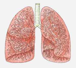
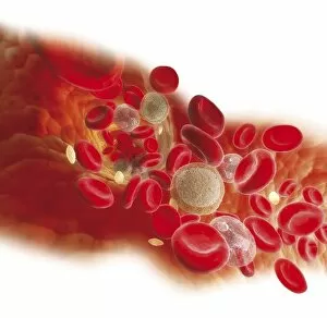
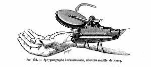
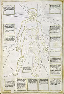
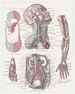
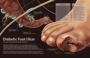
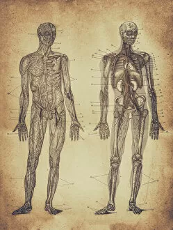
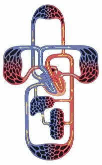
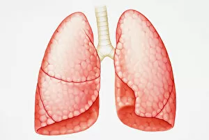
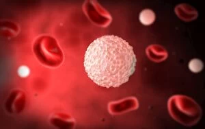
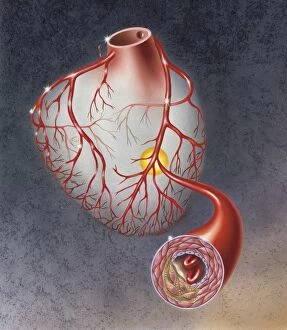
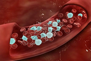
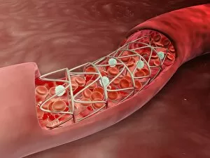
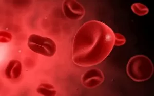
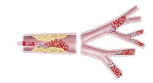
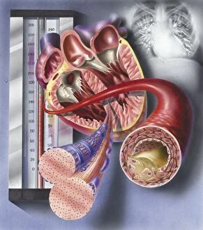
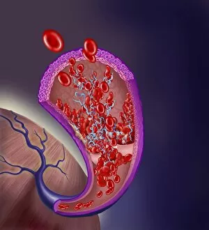
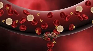
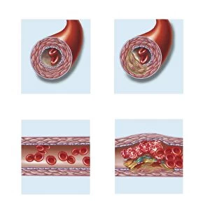
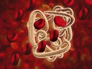
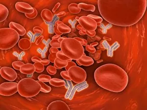
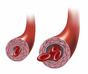
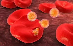
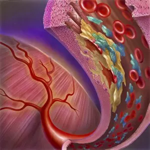
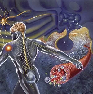
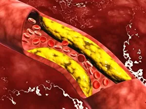
"Journey of Life: Exploring the Intricate Pathways of the Bloodstream" Step back in time to 1861, as a hand-coloured engraving unveils the marvels of the human circulatory system. This intricate diagram showcases the network that keeps us alive, revealing how blood flows through our veins and arteries. In another illustration from history, a 15th-century woodcut from Venice's Fasciculus medicinae takes us on a visual tour inside our lungs. The delicate balance between oxygen and carbon dioxide exchange is depicted with precision, highlighting the vital role played by blood supply in this process. Moving forward to 1874, an exquisite lithograph offers an anatomy lesson on the complexity of our bloodstream. Red and white blood cells dance within veins while platelets stand guard against potential harm—a mesmerizing sight that reminds us of life's fragility and resilience. Meanwhile, tragedy strikes in Macedonia during 1903 as Greek Orthodox wedding party members fall victim to Turkish aggression. A haunting print captures this somber event, serving as a stark reminder that even amidst chaos and violence, every heartbeat relies on a functioning bloodstream. Fast forward to vibrant hues—yellow ground contrasting with a red heart—an artistic representation symbolizing vitality and passion coursing through our veins. It serves as a vivid reminder that love fuels not only emotions but also sustains life itself. Delving deeper into medical knowledge, we encounter lithographs published in 1875 depicting fetal development alongside detailed histopathology studies of diabetic foot ulcers' pathophysiology. These images highlight both beginnings and challenges faced by humanity throughout its existence—each intricately tied to the wonders occurring within our bloodstream. A pair of human lungs emerges next—a testament to their crucial role in respiration—and prompts reflection on how they rely on efficient circulation for optimal function. Such interconnectedness underscores nature's remarkable design within each living organism.