
Bronchi Collection
Bronchi: The Vital Pathways of the Respiratory System The bronchi, a crucial component of our respiratory system, play a significant role in ensuring efficient breathing


All Professionally Made to Order for Quick Shipping
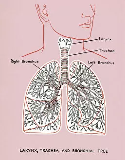
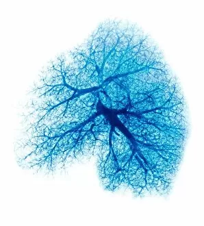
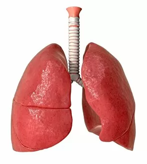
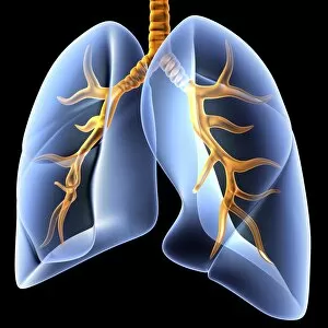
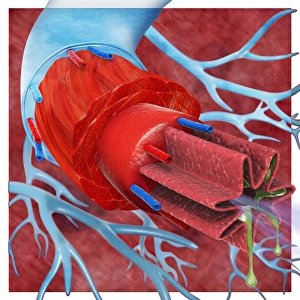
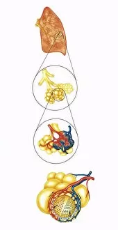
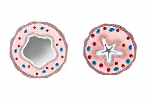
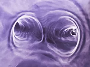
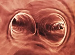
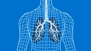
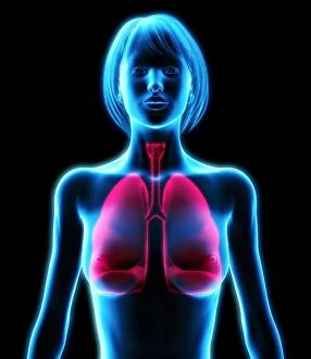
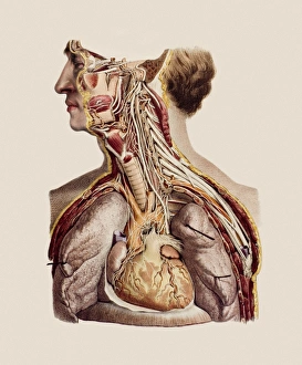
Bronchi: The Vital Pathways of the Respiratory System The bronchi, a crucial component of our respiratory system, play a significant role in ensuring efficient breathing. As depicted in the lung diagram and cross-section biomedical illustrations, these intricate air passages are intricately connected to the trachea and lungs. In human lungs, they branch out like delicate tree branches, transporting oxygen-rich air from the trachea into smaller airways called bronchioles. These blood vessels of the chest and neck supply vital nutrients to support their function. Anatomical artwork showcases how inflamed they are cause breathing difficulties during childhood. This condition is further illustrated through digital cross-sections where allergens such as pollen infiltrate both the trachea and bronchioles, triggering discomfort for individuals with allergies. For asthmatics, another digital illustration demonstrates how their trachea and bronchioles differ from those without this respiratory condition. Asthma causes constriction within these pathways leading to reduced airflow. Understanding how medications work is essential for managing respiratory ailments effectively. Biomedical illustrations reveal that before ingesting a bronchodilator drug, muscle contractions narrow the airway walls causing restricted breathing. However, after taking this medication, muscles relax allowing for increased airflow by widening these passageways. These captivating visuals provide invaluable insights into both healthy and compromised states of our respiratory system. Appreciating the complexity of our bronchi reminds us of their significance in maintaining optimal lung function while emphasizing proper care for overall well-being.