
Capsid Structure Collection
The capsid structure is a fascinating aspect of viral biology, playing a crucial role in protecting the genetic material and facilitating infection


All Professionally Made to Order for Quick Shipping
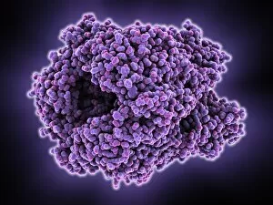
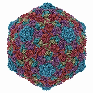
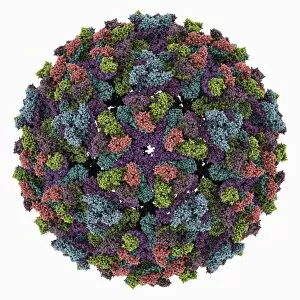
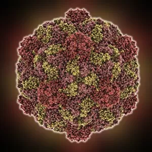
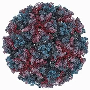
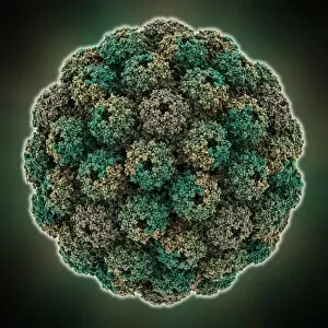
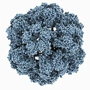
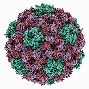
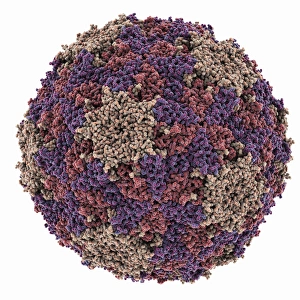
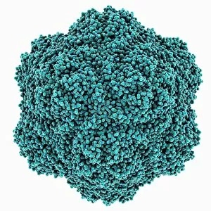
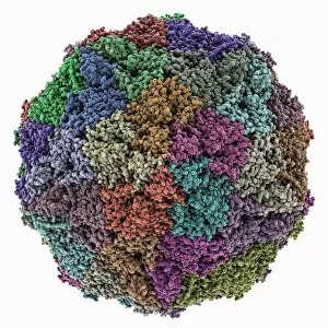
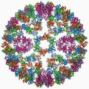
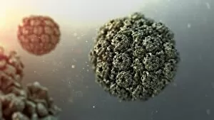
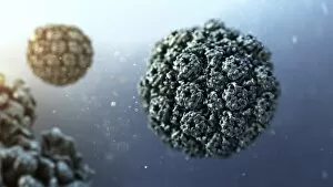
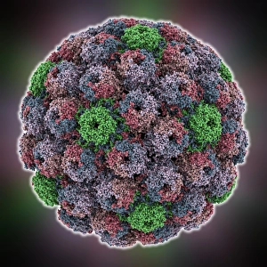
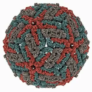
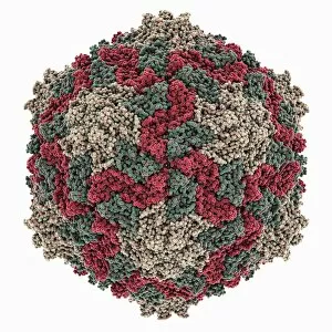
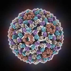
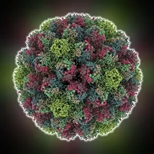
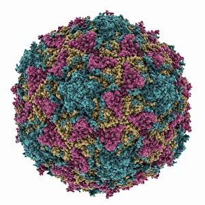
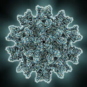
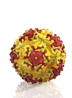
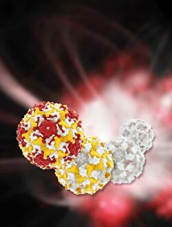
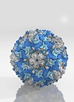
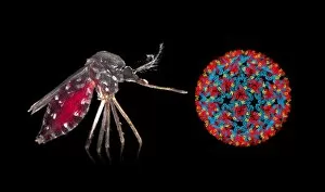
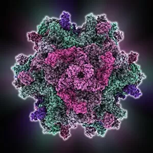
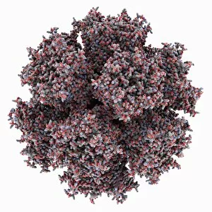
The capsid structure is a fascinating aspect of viral biology, playing a crucial role in protecting the genetic material and facilitating infection. Various viruses exhibit unique capsid structures, each with its own distinct features. One such example is the Adenovirus hexon protein, which forms the major component of the virus's capsid. Its intricate arrangement allows for efficient packaging of DNA within the virus particle. Another intriguing they are be found in the HK97 bacteriophage. This complex assembly consists of multiple proteins that come together to form an icosahedral shape, providing stability and protection to its genetic material. The Chikungunya virus also possesses a distinctive capsid structure that enables it to invade host cells efficiently. Its spherical shape contains numerous spikes protruding from its surface, aiding in attachment and entry into target cells. Similarly, Turnip yellow mosaic virus showcases a unique architecture with T-shaped protrusions on its surface. These play a vital role in recognizing specific receptors on host cells during infection. The Hepatitis B virus exhibits an icosahedral capsid structure composed of repeating subunits arranged symmetrically around a central axis. This molecular model provides valuable insights into how this virus evades immune responses and persists within infected individuals. Infectious bursal disease virus presents yet another captivating capsid structure characterized by elongated fibers extending from its surface. These fibers aid in receptor recognition and subsequent cell entry during infection. Sindbis virus boasts an elegant molecular model showcasing an icosahedral symmetry with prominent spikes decorating its outer shell. These spikes are essential for binding to host cell receptors and initiating viral replication processes. Murine polyomavirus displays a spherical capsid structure enclosing its genome tightly packed inside. The precise organization of proteins forming this protective shell ensures successful transmission between hosts. Brome mosaic virus exhibits an unusual rod-like morphology consisting of stacked discs forming helical arrays within the viral particle's interior space. This unique capsid structure enables efficient packaging of its RNA genome.