
Capsomere Collection
"Exploring the Intricate World of Capsomeres: A Glimpse into Viral Architecture" Capsomeres, the building blocks of viral capsids


All Professionally Made to Order for Quick Shipping
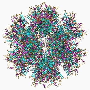
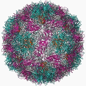
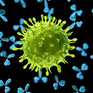
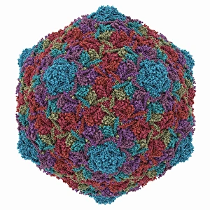
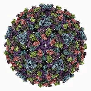
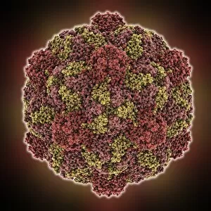
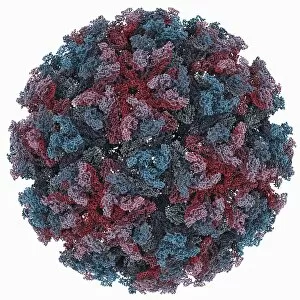
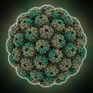
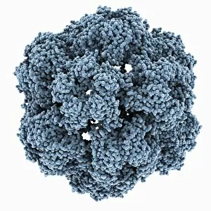
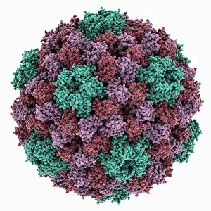
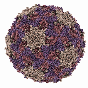
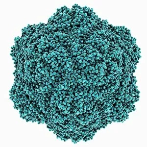
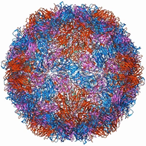
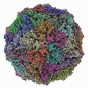
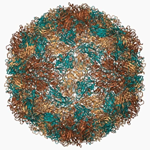
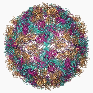
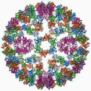
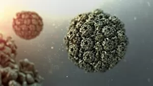
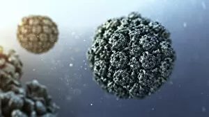
"Exploring the Intricate World of Capsomeres: A Glimpse into Viral Architecture" Capsomeres, the building blocks of viral capsids, are fascinating structures that play a crucial role in protecting and delivering viral genetic material. These tiny protein subunits come together to form the outer shell of viruses, giving them their unique shapes and enabling them to infect host cells. From the Adenovirus penton base protein F006/9542 to the Rhinovirus 16 capsid molecular model F006/9431, scientists have been studying these intricate arrangements to better understand virus-host interactions. Captivating artwork showcases antibodies attacking viruses like never before - F007/6623, F007/6624, and F007/6622 depict this battle between our immune system and invading pathogens. The HK97 bacteriophage capsid reveals yet another captivating example of how nature constructs protective shells. Its complex structure serves as a blueprint for potential therapeutic interventions against bacterial infections. But it's not just bacteria; plant viruses also possess remarkable capsids. The Chikungunya virus capsid and Turnip yellow mosaic virus capsid demonstrate nature's diversity in designing these protective layers across different species. Moving on to human pathogens, we encounter Hepatitis B virus with its distinctive molecular model showcasing its unique capsid architecture. Understanding this structure is vital for developing effective antiviral treatments against this persistent infection. Infectious bursal disease virus presents another intriguing case study where researchers investigate how specific changes in the capsid can enhance vaccine development strategies against avian diseases. Lastly, Sindbis virus captivates scientists with its molecular model revealing key insights into its replication cycle and potential targets for antiviral therapies. As we delve deeper into understanding these diverse viral architectures such as Murine polyomavirus capsid or any other yet-to-be-discovered variants, we unlock new opportunities for combating infectious diseases through targeted interventions and vaccine development.