
Capsomeres Collection
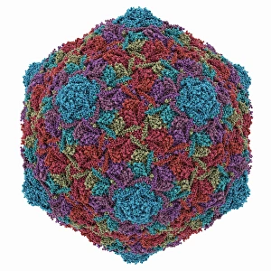
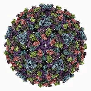
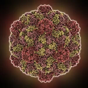
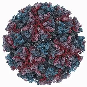
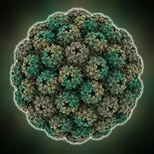
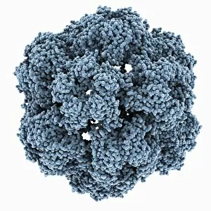
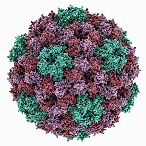
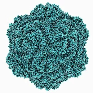
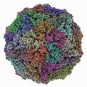
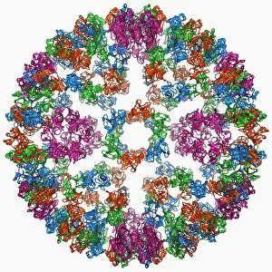
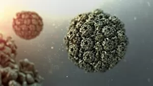
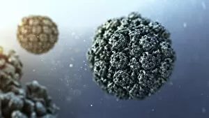
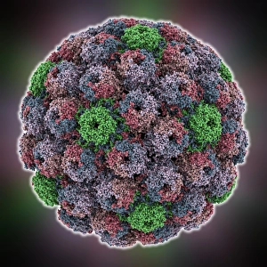
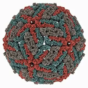
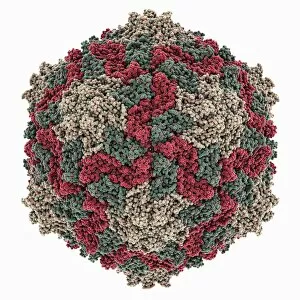
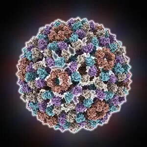
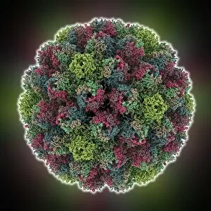
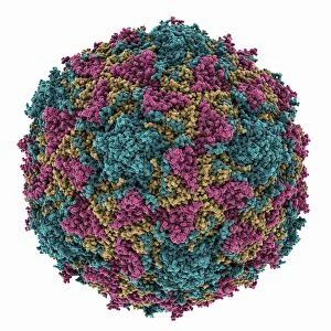
Capsomeres are the building blocks of viral capsids, which play a crucial role in protecting and delivering the genetic material of various viruses


All Professionally Made to Order for Quick Shipping
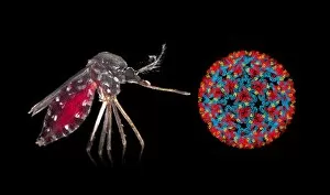
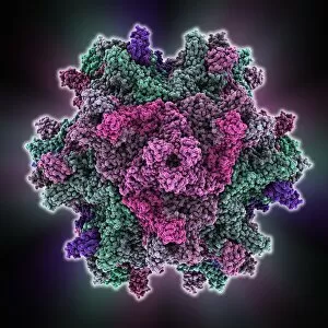
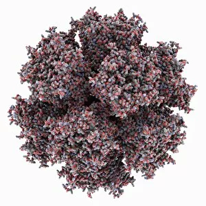
Capsomeres are the building blocks of viral capsids, which play a crucial role in protecting and delivering the genetic material of various viruses. These tiny protein subunits come together to form the outer shell of viral particles, providing structural integrity and facilitating infection. One example is the HK97 bacteriophage capsid, which encloses its DNA like a protective armor. Similarly, the Chikungunya virus capsid shields its RNA genome from host defenses as it invades human cells. The Turnip yellow mosaic virus capsid exhibits an intricate architecture that allows efficient packaging of its genetic material. Meanwhile, the Hepatitis B virus capsid boasts a molecular model that reveals its complex assembly process and potential targets for antiviral therapies. Infectious bursal disease virus relies on its distinctive capsid to invade immune cells in chickens, causing severe immunosuppression. The Sindbis virus also utilizes its unique molecular model to enter host cells and replicate within them. The Murine polyomavirus showcases another remarkable capsid structure involved in infecting rodents by targeting specific cell receptors. Similarly, Brome mosaic virus employs an elaborate arrangement to ensure successful replication within plant hosts. Semliki forest virus captivates scientists with its intricate symmetrical pattern formed by numerous identical proteins coming together harmoniously. On the other hand, Dengue virus presents a molecular model revealing how multiple copies of their distinctively shaped capsids interact with each other during infection. Lastly, Cowpea chlorotic mottle virus demonstrates how diverse viruses can employ different strategies when constructing their protective shells using individualized arrangements of protein subunits.