Fraud Blocker Skip to main content

Cell Biology Collection

"Exploring the Intricacies of Cell Biology: A Fascinating Journey into Cellular Life" Nerve and glial cells, captured through a mesmerizing light micrograph

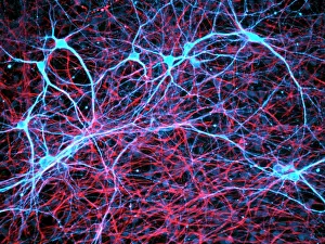
Background imageCell Biology Collection: Nerve and glial cells, light micrograph

Nerve and glial cells, light micrograph
Nerve and glial cells, fluorescence light micrograph. These are neural stem cells that have differentiated into neurons (nerve cells, blue) and glial cells (support cells, red)

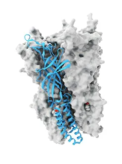
Background imageCell Biology Collection: Anaesthetic inhibiting an ion channel C015 / 6718

Anaesthetic inhibiting an ion channel C015 / 6718
Anaesthetic inhibiting an ion channel. Computer model showing the structure of propofol anaesthetic drug molecules (spheres)

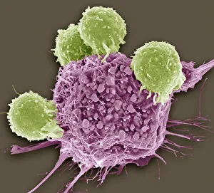
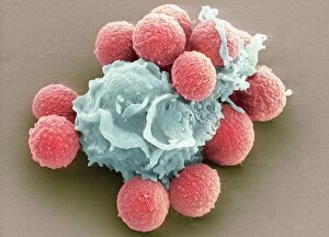
Background imageCell Biology Collection: T lymphocytes and cancer cell, SEM C001 / 1679

T lymphocytes and cancer cell, SEM C001 / 1679
T lymphocytes and cancer cell. Coloured scanning electron micrograph (SEM) of T lymphocyte cells (green) attached to a cancer cell

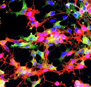
Background imageCell Biology Collection: Glial stem cell culture, light micrograph

Glial stem cell culture, light micrograph
Glial stem cell culture. Fluorescent light micrograph of glial stem cells producing the protein NG2 (red) as they mature. These stem cells can differentiate into several types of glial cells

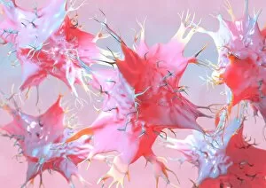
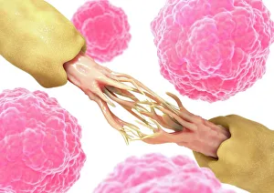
Background imageCell Biology Collection: Dendritic cells, artwork

Dendritic cells, artwork. Dendritic cells, a type of white blood cell, are called antigen presenting cells (APCs) and are found in the bodys tissues

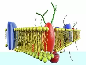
Background imageCell Biology Collection: Cell membrane, artwork C013 / 7467

Cell membrane, artwork C013 / 7467
Computer artwork of a cutaway view of the human cell membrane. The cell Membrane is a complex part of the cell that controls what can get in and out of the cell

Background imageCell Biology Collection: Pine pollen grains, light micrograph

Pine pollen grains, light micrograph
Pine pollen grains. Light micrograph of a section through winged pollen grains (microspores) from a pine (Pinus sp.) tree

Background imageCell Biology Collection: Pine stem, light micrograph

Pine stem, light micrograph
Pine stem. Light micrograph of a section through the stem of a pine (Pinus sp.) tree, showing xylem tissue. The xylem is made up of tracheid cells (light pink)

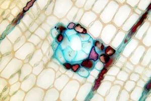
Background imageCell Biology Collection: Lime tree stem, light micrograph

Lime tree stem, light micrograph
Lime tree stem. Light micrograph of a section through the stem of a lime tree (Tilia europaea). The outer epidermis has been shed and replaced by a layer of cork (dark red)

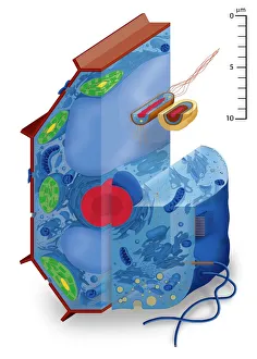
Background imageCell Biology Collection: Cell types, artwork

Cell types, artwork
Cell types. Cutaway artwork with a scale bar (upper right, in micrometres) showing the relative sizes of eukaryotic cells (those containing a nucleus) and prokaryotic cells (those lacking a nucleus)

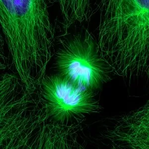
Background imageCell Biology Collection: Cell division, fluorescent micrograph

Cell division, fluorescent micrograph
Cell division. Immunofluorescent light micrograph of a human epithelial cell (centre) during the late anaphase stage of mitosis

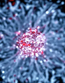
Background imageCell Biology Collection: Neural stem cell culture

Neural stem cell culture. Fluorescent light micrograph of a group of neural stem cells (neurosphere) in culture. Neural stem cells are able to differentiate into neurons (nerve cells)

Background imageCell Biology Collection: Glial cells, confocal light micrograph

Glial cells, confocal light micrograph
Glial cells. Confocal light micrograph of glial cells from the cerebellum of the brain. Glial cells are nervous system cells that provide structural support and protection for neurons (nerve cells)
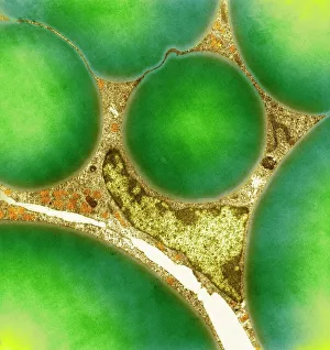
Background imageCell Biology Collection: Fat cells, TEM

Fat cells, TEM
Fat cells. Coloured transmission electron micrograph (TEM) of a section through fat (adipose) tissue, consisting of fat-storing cells (adipocytes, green) supported by connective tissue

Background imageCell Biology Collection: Dohle bodies in blood cell, micrograph

Dohle bodies in blood cell, micrograph
Dohle bodies in blood cell. Light micrograph of a neutrophil white blood cell (centre) with Dohle bodies. These are small inclusions within the cells cytoplasm

Background imageCell Biology Collection: Chloroplast structure, artwork

Chloroplast structure, artwork
Chloroplast structure. Artwork showing the internal structure of chloroplasts, the organelles in plant cells responsible for photosynthesis

Background imageCell Biology Collection: Water lily leaf, light micrograph

Water lily leaf, light micrograph
Water lily leaf. Light micrograph of a transverse section through the leaf of a water lily (Nympha sp.) plant. All aquatic plants (hydrophytes) have a similar structure

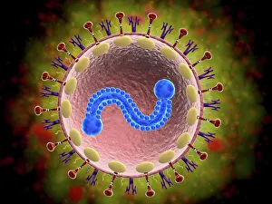
Background imageCell Biology Collection: Microscopic view of human respiratory syncytial virus

Microscopic view of human respiratory syncytial virus (RSV). RSV causes respiratory tract infection of the lungs and breathing passages

Background imageCell Biology Collection: Activated macrophage, SEM C015 / 6375

Activated macrophage, SEM C015 / 6375
Activated macrophage. Coloured scanning electron micrograph (SEM) of an active macrophage white blood cell. Macrophages are cells of the bodys immune system that are found in the tissues rather than

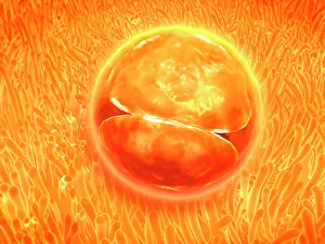
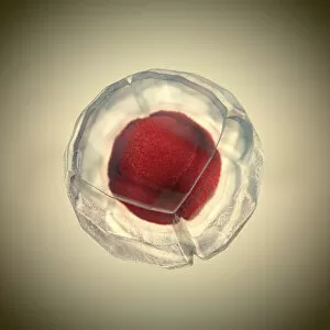
Background imageCell Biology Collection: Embryo development 24-36 hours after fertilization

Embryo development 24-36 hours after fertilization

Background imageCell Biology Collection: Plasma cells, TEM

Plasma cells, TEM
Plasma cells. Coloured transmission electron micrograph (TEM) of plasma cells. Plasma cells are mature B lymphocytes (white blood cells) that produce and secrete antibodies during an immune response

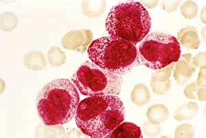
Background imageCell Biology Collection: Acute promyelocytic leukaemia, micrograph

Acute promyelocytic leukaemia, micrograph
Acute promyelocytic leukaemia. Light micrograph of blood cells from bone marrow in a case of acute promyelocytic leukaemia. Leukaemia is a cancer where certain blood cells form in excess

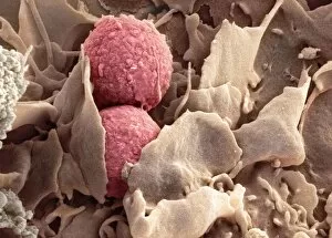
Background imageCell Biology Collection: Phagocytosis of fungal spores, SEM

Phagocytosis of fungal spores, SEM
Phagocytosis of a fungus spore. Coloured scanning electron micrograph (SEM) of a neutrophil (blue) phagocytosing (engulfing and destroying) spores from the fungus Aspergillus fumigatus (red)

Background imageCell Biology Collection: Cell division, fluorescent micrograph

Cell division, fluorescent micrograph
Cell division. Immunofluorescent light micrograph of a human epithelial cell (centre) during the interphase stage of mitosis
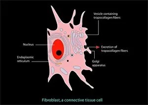
Background imageCell Biology Collection: Fibroblast cell, artwork

Fibroblast cell, artwork
Fibroblast cell. Computer artwork of a fibroblast excreting collagen fibres (tropocollagen). Fibroblasts are cells that produce connective tissue such as collagen (tropocollagen)

Background imageCell Biology Collection: Golgi apparatus, SEM

Golgi apparatus, SEM
Golgi apparatus, coloured scanning electron micrograph (SEM). Section through a liver cell showing its Golgi apparatus (grey), a membrane-bound organelle that modifies and packages proteins

Background imageCell Biology Collection: Ovarian follicle, light micrograph C016 / 0519

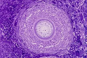
Ovarian follicle, light micrograph C016 / 0519
Ovarian follicle. Light micrograph of a section through a secondary ovarian follicle showing signs of maturation into the antral follicle class

Background imageCell Biology Collection: Eye muscle, TEM C014 / 1468

Eye muscle, TEM C014 / 1468
Eye muscle. Transmission electron micrograph (TEM) of a section through a striated muscle cell from the ciliary muscle of a human eye

Background imageCell Biology Collection: Purkinje nerve cell, TEM C014 / 0583

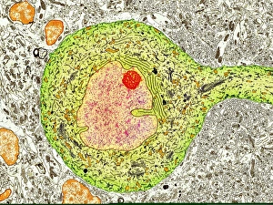
Purkinje nerve cell, TEM C014 / 0583
Purkinje nerve cell. Transmission electron micrograph (TEM) of a purkinje nerve cell (green) from the cerebellum of the brain, showing the cell body (centre) and its primary dendrite (cell process)

Background imageCell Biology Collection: Lymphocyte white blood cells, artwork

Lymphocyte white blood cells, artwork
Lymphocyte white blood cells. Computer artwork of lymphocyte white blood cells, showing their nuclei (red, centre). Lymphocytes, like all white blood cells, are part of the immune system

Background imageCell Biology Collection: Intestinal microvilli, TEM

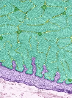
Intestinal microvilli, TEM
Intestinal microvilli, coloured transmission electron micrograph (TEM). The microvilli are finger-like projections (seen here in circular cross-section)

Background imageCell Biology Collection: Nerve damage and stem cells, artwork

Nerve damage and stem cells, artwork
Nerve damage and stem cells, computer artwork. Stem cells are undifferentiated cells that can produce other types of cell when they divide

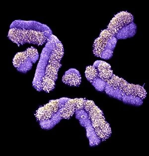
Background imageCell Biology Collection: Human chromosomes, SEM C013 / 5002

Human chromosomes, SEM C013 / 5002
Human chromosomes. Coloured scanning electron micrograph (SEM) of human chromosomes prepared with the harlequin staining technique

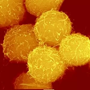
Background imageCell Biology Collection: Haematopoietic stem cells, SEM C013 / 5009

Haematopoietic stem cells, SEM C013 / 5009
Haematopoietic stem cells, coloured scanning electron micrograph (SEM). Stem cells can differentiate into any other cell type

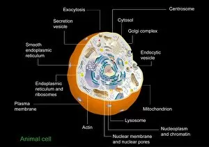
Background imageCell Biology Collection: Animal cell structure

Animal cell structure. Artwork of a section through an animal cell. At lower centre is the nucleus, which contains the cells genetic information in the form of DNA (deoxyribonucleic acid)

Background imageCell Biology Collection: Cell, illustration

Cell, illustration
3d illustration of cell division, cell membrane and a splitting red nucleus

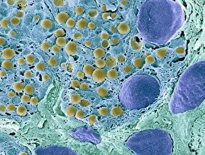
Background imageCell Biology Collection: Pancreas tissue, colored scanning electron micrograph

Pancreas tissue, colored scanning electron micrograph
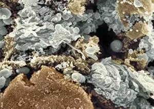
Pancreas tissue. Coloured scanning electron micrograph (SEM) of fractured pancreas tissue. Seen here are zymogen granules (yellow) and cell nuclei (purple)

Background imageCell Biology Collection: Microscopic view of animal cell

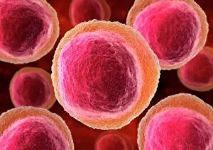
Microscopic view of animal cell

Background imageCell Biology Collection: A female nude from the waist up with a doctors hands conducting a clinical breast

A female nude from the waist up with a doctors hands conducting a clinical breast examination

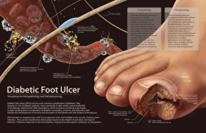
Background imageCell Biology Collection: Histopathology and pathophysiology of diabetic food ulcers

Histopathology and pathophysiology of diabetic food ulcers

Background imageCell Biology Collection: TFAM transcription factor bound to DNA C015 / 7059

TFAM transcription factor bound to DNA C015 / 7059
TFAM transcription factor bound to DNA, molecular model. Human mitochondrial transcription factor A (TFAM, green) bound to a strand of DNA (deoxyribonucleic acid, blue and pink)

Background imageCell Biology Collection: Phagocytosis of fungus spores, SEM

Phagocytosis of fungus spores, SEM
Phagocytosis of fungus spores. Coloured scanning electron micrograph (SEM) of spores from the fungus Aspergillus fumigatus (red) being phagocytosed (engulfed and destroyed) by a dendritic cell (brown)

Background imageCell Biology Collection: Anton van Leeuwenhoek, caricature

Anton van Leeuwenhoek, caricature
Anton van Leeuwenhoek. Caricature of the Dutch microbiologist Anton van Leeuwenhoek (1632-1723), looking through a magnifying glass to represent his discoveries in microbiology

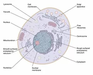
Background imageCell Biology Collection: Animal cell anatomy, diagram

Animal cell anatomy, diagram
Animal cell anatomy. Diagram showing the internal and external anatomy of an animal cell

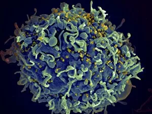
Background imageCell Biology Collection: Scanning electron micrograph of HIV particles infecting a human H9 T cell

Scanning electron micrograph of HIV particles infecting a human H9 T cell

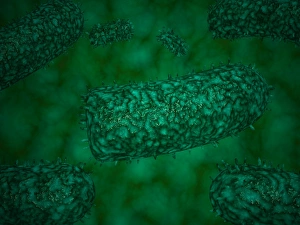
Background imageCell Biology Collection: Coxiella burnetii, the bacteria that causes Q Fever

Coxiella burnetii, the bacteria that causes Q Fever. A dry fracture of a Vero cell exposing the contents of a vacuole where Coxiella burnetii are busy growing

Background imageCell Biology Collection: Conceptual image of platelet, red blood cell and white blood cell

Conceptual image of platelet, red blood cell and white blood cell

Background imageCell Biology Collection: Stylized rabies virus particles

Stylized rabies virus particles, the cause of the viral neuroinvasive disease acute encephalitis


All Professionally Made to Order for Quick Shipping

"Exploring the Intricacies of Cell Biology: A Fascinating Journey into Cellular Life" Nerve and glial cells, captured through a mesmerizing light micrograph, reveal the intricate network that supports our nervous system. T lymphocytes battling cancer cells - witness this epic battle in stunning detail with SEM C001 / 1679 imagery. Anaesthetic inhibiting an ion channel - delve into the world of cellular communication as we uncover how anaesthetics affect these vital channels (C015 / 6718). Glial cells take center stage in a captivating confocal light micrograph, showcasing their crucial role in supporting neurons. Witness the beauty of glial stem cell culture under a microscope; it's like exploring an enchanting galaxy within our own bodies (light micrograph). Dendritic cells come to life through exquisite artwork, highlighting their pivotal role in immune response and defense mechanisms. Pine pollen grains unveil their intricate structure under a light microscope – nature's tiny marvels never cease to amaze us. Dive deep into the inner workings of pine stems with a captivating light micrograph – discover the building blocks that support these majestic trees. Explore the hidden wonders of lime tree stems through a mesmerizing light micrograph – witness nature's architectural masterpiece up close. Uncover various cell types brought to life through breathtaking artwork – each one playing its unique part in maintaining life's delicate balance. Immerse yourself in neural stem cell culture and witness firsthand how these remarkable cells hold immense potential for regenerative medicine. The artistic portrayal of cell membranes (C013 / 7467) invites you to ponder upon their crucial role as gatekeepers controlling molecular traffic within living organisms. Embark on this awe-inspiring journey into cell biology and unlock the secrets that shape all living beings from within.