
Cell Death Collection
"Unveiling the Enigma of Cell Death: A Journey into the Intricacies of Mitochondria" In a world unseen by the naked eye


All Professionally Made to Order for Quick Shipping
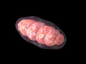
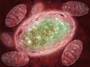
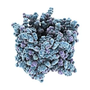
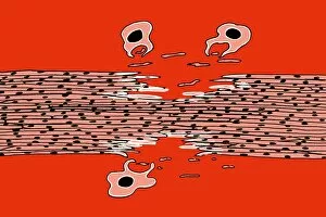
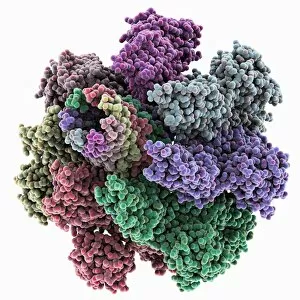
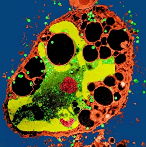
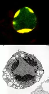
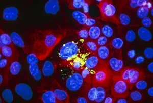
"Unveiling the Enigma of Cell Death: A Journey into the Intricacies of Mitochondria" In a world unseen by the naked eye, lies a realm where life and death intertwine - within the intricate structures of mitochondria. This conceptual image captures their enigmatic essence, inviting us to delve deeper into their microscopic view. Mitochondria, often referred to as the powerhouses of cells, hold secrets that transcend mere energy production. They possess an intrinsic role in cell death - a phenomenon essential for maintaining balance in our biological systems. This captivating conceptual image portrays their significance in this delicate dance between life and demise. Peering through the lens of a microscope reveals another dimension within these tiny organelles. The microscopic view unravels hidden details, showcasing their complex architecture and unveiling clues about cellular fate. But what drives this mysterious process? Ischaemic bowel, depicted here in a light micrograph, serves as an example where cell death plays a crucial role. Understanding how mitochondria contribute to such conditions can pave new paths towards therapeutic interventions. Beyond human physiology lies another realm where E. coli bacteria thrive. These SEM images capture their existence on a different scale but remind us that even at this level, cell death remains an integral part of life's tapestry. Exploring its intricacies within bacterial realms may provide insights applicable across diverse organisms. As we journey further into understanding cell death's complexities, let us embrace both conceptual and microscopic views with awe and curiosity. For it is through unraveling these mysteries that we unlock potential avenues for medical advancements and gain profound insights into the very essence of life itself.