Fraud Blocker Skip to main content

Cell Collection (page 100)

"Exploring the Intricacies of Life: Unveiling the Wonders of Cells" Delving into the microscopic world

Background imageCell Collection: Bacteria shapes, artwork

Bacteria shapes, artwork
Bacterial shapes. Computer artwork showing the appearance of various different types of bacteria
Background imageCell Collection: Alzheimers disease, computer artwork

Alzheimers disease, computer artwork
3D computer artwork depicting senile amyloid plaque (brown) in the brain, which kill surrounding neurons (blue). Large numbers of senile plaques are characteristic features of Alzheimers disease

Background imageCell Collection: Bacterial replication, artwork

Bacterial replication, artwork
Bacterial replication. Computer artwork showing a single bacterium dividing to form two identical daughter cells. This process is a form of asexual reproduction and is known as binary fission

Background imageCell Collection: Protein, microtubules and cell, artwork

Protein, microtubules and cell, artwork
3D computer artwork of a protein attached to microtubules, transporting vesicles and other important parts of the cell. Microtubules are polymers of the protein tubulin

Background imageCell Collection: Photosynthesis, diagram

Photosynthesis, diagram
Photosynthesis. Diagram showing the inputs and outputs (coloured arrows) involved in photosynthesis in the leaf of a plant

Background imageCell Collection: Retinal rod cell anatomy, diagram

Retinal rod cell anatomy, diagram

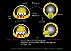
Background imageCell Collection: Vestibular ciliated cells, diagram

Vestibular ciliated cells, diagram
Vestibular ciliated cells. Diagram of the anatomical structure and function of the vestibular ciliated cells found in the inner ear that help sense and control of balance

Background imageCell Collection: Mirror neurons, conceptual image

Mirror neurons, conceptual image
Mirror neurons, conceptual computer artwork. Nerve cells, or neurons, and a human eye reflected on a shiny surface representing a mirror neuron cell. Mirror neurons are found in the brain

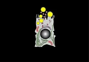
Background imageCell Collection: Milk-producing cell, artwork

Milk-producing cell, artwork
Milk-producing cell. Artwork of the anatomical structure of a lacteal (milk-producing) cell. Normal cell organelles include the nucleus (black, centre), the golgi apparatus (white)

Background imageCell Collection: ATP sulfurylase molecule

ATP sulfurylase molecule. Computer model showing the structure of an ATP sulfurylase (ATPS) enzyme. ATPS catalyses the primary step of sulphate activation within cells

Background imageCell Collection: Vestibular ciliated cells, artwork

Vestibular ciliated cells, artwork
Vestibular ciliated cells. Artwork of the anatomical structure and function of the vestibular ciliated cells found in the inner ear that help sense and control of balance

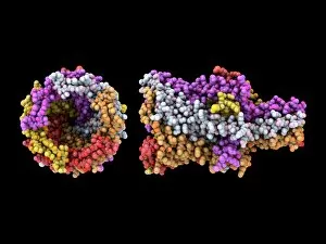
Background imageCell Collection: Prokaryotic mechanosensitive ion channel

Prokaryotic mechanosensitive ion channel molecule. Computer model showing two views of the molecular structure of a Mechanosensitive Channel of Large Conductance (MscL)

Background imageCell Collection: Acrosome reaction, artwork

Acrosome reaction, artwork
Acrosome reaction. Computer artwork showing the process by which an acrosome from a spermatozoon (sperm cell) binds to an ovum (egg cell, bottom). Each spermatozoon has an acrosome in its tip

Background imageCell Collection: Solar power plant, Nevada, USA

Solar power plant, Nevada, USA
Solar power plant. Rows of photovoltaic arrays at the Nellis Solar Power Plant, Nellis Air Force Base, Nevada, USA. This 14 megawatt energy system generates in excess of 25 million kilowatt-hours of

Background imageCell Collection: Retinal rod cell anatomy, artwork

Retinal rod cell anatomy, artwork
Retinal rod cell anatomy. Artwork of the external (left) and internal (right) anatomical structure of a retinal cell. These cells are found in the retina that lines the back of the eyes

Background imageCell Collection: Nerve cell with electrical sparks

Nerve cell with electrical sparks
Nerve cell. Conceptual computer artwork of a nerve cell, also called a neuron emitting electrical sparks. Neurons are responsible for passing information around the central nervous system (CNS)

Background imageCell Collection: Milk-producing cell, diagram

Milk-producing cell, diagram
Milk-producing cell. Diagram of the anatomical structure of a lacteal (milk-producing) cell. Normal cell organelles include the nucleus (black, centre), the golgi apparatus (white)

Background imageCell Collection: Mirror neuron, conceptual image

Mirror neuron, conceptual image
Mirror neuron, conceptual computer artwork. Nerve cell, or neuron, reflected in water, representing a mirror neuron cell. Mirror neurons are found in the brain

Background imageCell Collection: Animal cell anatomy, artwork

Animal cell anatomy, artwork
Animal cell anatomy. Artwork showing the internal and external anatomy of an animal cell

Background imageCell Collection: Macrophage engulfing pathogen, artwork

Macrophage engulfing pathogen, artwork
Macrophage engulfing pathogen. Artwork of a macrophage cell (pink) engulfing a pathogen (green), a process known as phagocytosis. The pathogen will be destroyed once it has been engulfed

Background imageCell Collection: Insect anatomy, artwork

Insect anatomy, artwork
Insect anatomy. Computer artwork of a cross-section through the body of a typical Insect showing the main structures and organs. For the labelled diagram see image: C008/8724

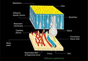
Background imageCell Collection: Olfactory epithelium, artwork

Olfactory epithelium, artwork
Olfactory epithelium. Computer artwork showing the structure of the specialised layer of tissue that lines the inside of the nasal cavity and is involved in smell

Background imageCell Collection: Activated platelets, artwork

Activated platelets, artwork
Activated platelets. Artwork of platelets (thrombocytes) that have been activated. Platelets are part of the blood. When a blood vessel is damaged

Background imageCell Collection: Macrophage engulfing pathogens, artwork

Macrophage engulfing pathogens, artwork
Macrophage engulfing pathogens. Artwork of a macrophage cell (pink) engulfing pathogens (green), a process known as phagocytosis. The pathogens will be destroyed once they have been engulfed

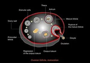
Background imageCell Collection: Ovarian cycle, artwork

Ovarian cycle, artwork
Ovarian cycle, computer artwork. Once sexual maturity is reached up to 20 oocytes start developing into eggs (ovums) every menstrual cycle

Background imageCell Collection: Human fertilisation, artwork

Human fertilisation, artwork
Human fertilization. Artwork of a single sperm cell (spermatozoon) attempting to fertilise a human egg cell (ovum, round, lower right). The sperm cell consists of a head section and a tail section

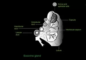
Background imageCell Collection: Exocrine gland, artwork

Exocrine gland, artwork
Exocrine gland. Computer artwork of a section through an exocrine gland. Exocrine glands secrete their products through ducts on to epithelial surfaces

Background imageCell Collection: mRNA leaving the nucleus, artwork

mRNA leaving the nucleus, artwork. mRNA (messenger ribonucleic acid, orange) is the intermediary molecule between DNA (deoxyribonucleic acid) and its protein product

Background imageCell Collection: Ovarian follicle maturation, artwork

Ovarian follicle maturation, artwork
Ovarian follicle maturation. Computer artwork showing the beginning of the follicular phase of ovarian follicle maturation. At bottom left is the primary follicle stage

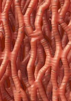
Background imageCell Collection: Cardiac muscle, artwork

Cardiac muscle, artwork
Cardiac muscle. Computer artwork of cardiac muscle fibres. Cardiac muscle is under subconscious control and continuously contracts to pump blood around the body without tiring

Background imageCell Collection: Blood cells, SEM

Blood cells, SEM
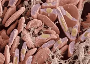
Blood cells. Coloured scanning electron micrograph (SEM) of a human red blood cell (erythrocyte, red) and a white blood cell (leucocyte, blue). These are the two main cell components of blood

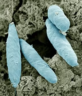
Background imageCell Collection: Euglena flagellate protozoans, SEM

Euglena flagellate protozoans, SEM
Euglena flagellate protozoans. Scanning electron micrograph (SEM) of Euglena sp. flagellate protozoans. Euglena sp. is a freshwater unicellular (single-celled)

Background imageCell Collection: Stem cell, artwork

Stem cell, artwork
Stem cell with a glowing nucleus, computer artwork. A stem cell is an undifferentiated cell that can produce other types of cell when it divides

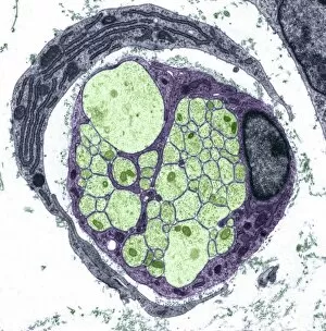
Background imageCell Collection: Nerve fibre, TEM

Nerve fibre, TEM
Nerve fibre. Coloured transmission electron micrograph (TEM) of a section through a non-myelinated peripheral nerve fibre

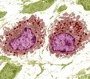
Background imageCell Collection: Macrophage cells, TEM

Macrophage cells, TEM
Macrophage cells, coloured transmission electron micrograph (TEM). The cell nuclei are purple. Mitochondria (dark pink ovals) in the cytoplasm produce energy for the cell

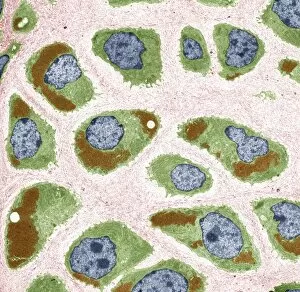
Background imageCell Collection: Cartilage cells, TEM

Cartilage cells, TEM
Cartilage cells. Coloured transmission electron micrograph (TEM) of a section through chondrocytes from nasal hyaline cartilage

Background imageCell Collection: Bird red blood cells, SEM

Bird red blood cells, SEM
Bird red blood cells. Coloured scanning electron micrograph (SEM) of fractured red blood cells (erythrocytes, red) from avian heart tissue

Background imageCell Collection: Marquis de Condorcets last meal

Marquis de Condorcets last meal, artwork. French politician, mathematician and philosopher Marie Jean Antoine Nicolas de Caritat, Marquis de Condorcet

Background imageCell Collection: Abdominal fat tissue, SEM

Abdominal fat tissue, SEM
Abdominal fat tissue, coloured scanning electron micrograph (SEM). Strands of connective tissue are seen running across the fat cells (adipocytes)

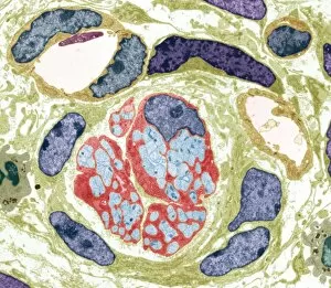
Background imageCell Collection: Unmyelinated nerve bundle, TEM

Unmyelinated nerve bundle, TEM
Unmyelinated nerve bundle. Coloured transmission electron micrograph (TEM) of a section through a group of unmylinated nerve fibres


All Professionally Made to Order for Quick Shipping

"Exploring the Intricacies of Life: Unveiling the Wonders of Cells" Delving into the microscopic world, a histological diagram of a mammalian retina reveals the intricate structure and organization of cells that enable us to perceive light. The cerebellum tissue, captured in a light micrograph, showcases its complex network of cells responsible for coordinating movement and balance. Intriguingly, even philosophers find solace in contemplating cellular existence. "Philosopher in Meditation, " an oil painting from 1632, reminds us that pondering life's mysteries often leads us back to our fundamental building blocks – cells. Zooming further into this realm, nerve and glial cells come alive under the lens. This captivating light micrograph highlights their vital role in transmitting signals throughout our nervous system. Meanwhile, a lavender pollen grain captured through scanning electron microscopy (SEM) displays nature's exquisite design at the cellular level. The battle against diseases takes center stage as T lymphocytes confront cancer cells in another SEM image. Science and evolution intertwine as we witness these tiny warriors fighting for survival within our bodies. Artistic expressions also shed light on cell significance; "The Death of Socrates, " painted in 1787, serves as a poignant reminder that even great minds are ultimately composed of countless individual cells. Santiago Ramon y Cajal's meticulous drawing from 1894 unveils various cell types within the mammalian cerebellum. His work not only represents scientific progress but also exemplifies how art can aid understanding by visually capturing complexity. Stepping away from biology momentarily, we encounter Rolls Royce/Snecma Olympus 593 Mk602 engine undergoing testing within a controlled environment called a test cell. Here too, precision engineering relies on understanding cellular mechanics to achieve optimal performance. Returning to biological marvels - HeLa cells take center stage under yet another microscope lens. These immortalized human cells have revolutionized medical research, paving the way for countless breakthroughs.