
Cerebrospinal Fluid Collection
"Cerebrospinal fluid: The Lifeline of the Brain" Cerebrospinal fluid (CSF) is a vital component that plays an essential role in protecting and nourishing our brain


All Professionally Made to Order for Quick Shipping
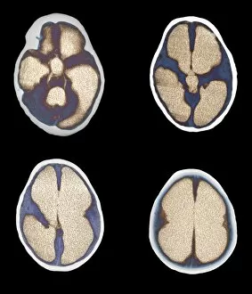
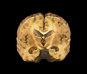
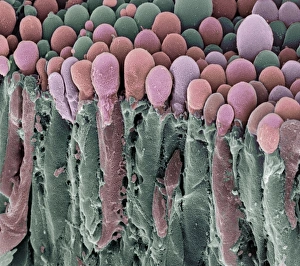
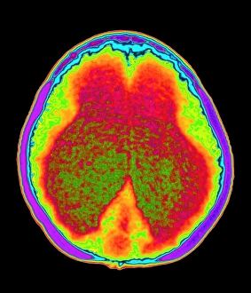
"Cerebrospinal fluid: The Lifeline of the Brain" Cerebrospinal fluid (CSF) is a vital component that plays an essential role in protecting and nourishing our brain. This clear, colorless liquid acts as a cushioning agent by enveloping the brain lining, safeguarding it from potential damage. Through scanning electron microscopy (SEM), we can observe fascinating images of fallopian tube cells. These microscopic structures resemble intricate networks, highlighting the complexity of CSF's origin within the ventricles of the brain. In cases like Spina Bifida or Meningocele, where protective covering around the spinal cord protrudes through malformed vertebrae, sacs filled with it can formed. A digital illustration depicts this condition vividly, emphasizing how crucial CSF management becomes for affected individuals. Biomedical illustrations exhibit cross-sections of cerebral shunts inserted into young patients' brains to remove excess cerebrospinal fluid. These life-saving devices consist of valves and tubes that redirect CSF flow towards the stomach for proper drainage. X-ray images showcase ventricular shunts used to alleviate conditions such as hydrocephalus—a buildup within cavities called ventricles—by diverting excessive fluids away from critical areas. Multiple X-rays emphasize their significance in maintaining normal intracranial pressure levels effectively. Amidst these medical interventions lies a healthy brain captured through MRI scans—an image revealing no abnormalities or irregularities. It serves as a reminder that cerebrospinal fluid's optimal circulation ensures our brains function at their best capacity. Cerebrospinal fluid acts as a lifeline for our brains—protective yet dynamic in its role. From SEM images showcasing cellular intricacies to biomedical illustrations depicting life-saving procedures involving shunting systems and diagnostic tools like X-rays and MRIs—the significance of understanding CSF cannot be overstated, and is through this understanding that we can appreciate the delicate balance required