Fraud Blocker Skip to main content

Chest Collection (page 76)

"The Chest: A Canvas of Stories and Secrets" Tattooed masterpiece by Mr

Background imageChest Collection: Radiologists examining an X-ray

Radiologists examining an X-ray
MODEL RELEASED. Radiologists studying an X-ray of a patients chest

Background imageChest Collection: Boy posing

Boy posing
MODEL RELEASED. Boy posing. He is six years old

Background imageChest Collection: Chest X-ray

Chest X-ray
MODEL RELEASED. Chest X-ray

Background imageChest Collection: Female torso, artwork

Female torso, artwork
Female torso. Computer artwork of a healthy womans upper body

Background imageChest Collection: Ribcage, computer artwork

Ribcage, computer artwork
Ribcage. Computer artwork of the pectoral girdle and ribcage as seen from the front

Background imageChest Collection: Human internal organs, diagram

Human internal organs, diagram
Human internal organs. Diagram showing the human internal organs in the head and torso. In the head, the organs and cavities shown are: the brain (encephalon), the cerebellum (part of the brain)

Background imageChest Collection: Stent to treat aortic aneurysm, CT scan

Stent to treat aortic aneurysm, CT scan
Coloured computed tomography (CT) angiography scan of the chest in a 55 year old diabetic patient showing three stents (endoprostheses)

Background imageChest Collection: Human internal organs, artwork

Human internal organs, artwork
Human internal organs. Artwork showing the human internal organs in the head and torso. In the head, the organs and cavities shown are: the brain (encephalon), the cerebellum (part of the brain)

Background imageChest Collection: Collapse (atelectasis) of lung, X-ray

Collapse (atelectasis) of lung, X-ray
Coloured X-ray (front view) of the chest in a 58 year old patient showing collapse (atelectasis) of part of the upper lobe of a lung (orange, at left)

Background imageChest Collection: Mechanics of respiration, artwork

Mechanics of respiration, artwork
Mechanics of respiration. Cutaway artworks showing the mechanics of breathing in (left) and breathing out (right). Arrows show: the movement of air in and out of the nose

Background imageChest Collection: Chest X-ray imaging, composite image

Chest X-ray imaging, composite image. At left a ceiling-mounted X-ray machine is being used to X-ray a patients chest. The artwork of the patient (at right)

Background imageChest Collection: Placing of electrodes for ECG, artwork

Placing of electrodes for ECG, artwork
Placing of electrodes for electrocardiogram (ECG), artwork. This diagram shows where the six electrodes attached to the chest should be placed. One electrode is also placed on each arm and leg

Background imageChest Collection: Healthy chest X-ray of a young child

Healthy chest X-ray of a young child
Coloured X-ray of the healthy chest in a two year old boy, showing the heart (pink) and lungs (dark) enclosed by the ribcage

Background imageChest Collection: Tuberculosis lymphadenopathy, X-ray

Tuberculosis lymphadenopathy, X-ray
Coloured X-ray (front view) of the chest in a 14 year old male patient, showing inflamed lymph nodes (lymphadenopathy, yellow, at centre right and left) caused by tuberculosis (TB)

Background imageChest Collection: Pulmonary consolidation, X-ray

Pulmonary consolidation, X-ray
Pulmonary consolidation. X-ray showing that the upper lobe of a patients left lung has become consolidated (filled with pus) due to pneumonia

Background imageChest Collection: Old and new tuberculosis, X-ray

Old and new tuberculosis, X-ray
Coloured X-ray (front view) of the chest in a 50 year old male patient, showing fibrosis in the lungs from old tuberculosis (more fibrous scarring, orange)

Background imageChest Collection: ECG electrode placement, artwork

ECG electrode placement, artwork
ECG electrode placement. Computer artwork showing the location of the 10 electrodes needed to perform a 12 lead electrocardiogram (ECG) test

Background imageChest Collection: Vascular anatomy, historical artwork

Vascular anatomy, historical artwork. 19th Century hand coloured lithographic print showing the arteries (red) and muscle structure (brown) of the human neck and chest

Background imageChest Collection: Heart and lungs, historical illustration

Heart and lungs, historical illustration. 19th Century hand coloured lithographic print showing the lungs (left and right) situated either side of the heart (lower centre)

Background imageChest Collection: Human arteries, 19th Century illustration

Human arteries, 19th Century illustration. Historical hand coloured lithographic print showing the arteries (red) and muscle structure (brown) of the human torso

Background imageChest Collection: Chest anatomy, 19th Century illustration

Chest anatomy, 19th Century illustration. Historical hand coloured lithographic print showing the lungs (centre) in the chest, larynx (voicebox, white, upper centre) in the throat

Background imageChest Collection: Thoracic lymphatic system, artwork

Thoracic lymphatic system, artwork
Thoracic lymphatic system. Artwork of a profile view of the thorax showing the lymphatic system and lymph nodes (red and white ovals). The lymphatic system is part of the immune system

Background imageChest Collection: Respiratory anatomy, 19th Century artwork

Respiratory anatomy, 19th Century artwork. Historical hand coloured lithographic print showing the trachea (wind pipe, vertical) running down from the larynx (voicebox)

Background imageChest Collection: Thorax anatomy, artwork

Thorax anatomy, artwork
Thorax anatomy. Computer artwork showing the bones, muscles, blood vessels and nerves of the thorax. At top right and left are the subclavian arteries (red) and veins (blue). Nerves are yellow

Background imageChest Collection: Ribcage and blood vessels, 3D CT scan

Ribcage and blood vessels, 3D CT scan
Coloured three-dimensional (3D) Computed Tomography (CT) scan in front view of the chest in a patient showing the bones of the ribcage (ribs) which protect the heart (at centre) and its blood vessels

Background imageChest Collection: Female body, artwork

Female body, artwork
Female body. Computer artwork showing the internal structure and organs of the female body

Background imageChest Collection: Abdominal air pockets, X-ray

Abdominal air pockets, X-ray

Background imageChest Collection: Emphysema, 3D CT scan

Emphysema, 3D CT scan

Background imageChest Collection: Aortic aneurysm, X-ray

Aortic aneurysm, X-ray
Aortic aneurysm. Coloured X-ray of the chest of a 68-year-old woman with an aortic aneurysm. An aneurysm is a swelling of an artery where the wall has weakened, here on the arch where the aorta

Background imageChest Collection: Fibrosing alveolitis, X-ray

Fibrosing alveolitis, X-ray
Fibrosing alveolitis. Coloured X-ray of the chest of an 82-year-old man with fibrosing alveolitis. Also known as idiopathic pulmonary fibrosis and cryptogenic fibrosing alveolitis

Background imageChest Collection: Respiratory nerves, 1844 artwork

Respiratory nerves, 1844 artwork
Respiratory nerves. These anatomical artworks are figures 2-3, plate 49, volume 3 (1844) of Traite complet de l anatomie de l homme (1831-1854)

Background imageChest Collection: Pneumonia, X-ray

Pneumonia, X-ray

Background imageChest Collection: Ribcage and heart, 3D CT scan

Ribcage and heart, 3D CT scan
Coloured three-dimensional (3D) Computed Tomography (CT) scan in side view of the chest in a patient showing the bones of the ribcage (ribs) which protect the heart (at centre) and its blood vessels

Background imageChest Collection: Bronchus and bronchioles, artwork

Bronchus and bronchioles, artwork
Bronchus and bronchioles. Artwork of the anatomical structure of the bronchi and their bronchioles, the main aspects of the respiratory system

Background imageChest Collection: Male vascular system, artwork

Male vascular system, artwork
Human male anatomy. Artwork showing the anatomy of a healthy adult male. The bones are brown and red, the brain (top) is pink and brown, and the heart (upper centre, behind the ribcage) is pink

Background imageChest Collection: Blood vessels and kidneys, artwork

Blood vessels and kidneys, artwork
Blood vessels and kidneys. Computer artwork of a man with healthy blood vessels and kidneys. The kidneys are at lower centre


All Professionally Made to Order for Quick Shipping

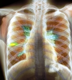
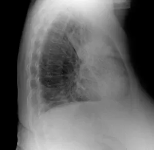
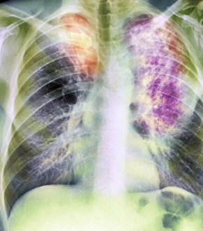
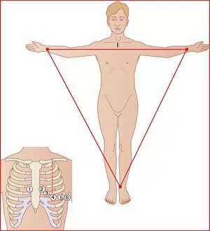
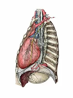
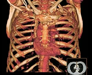
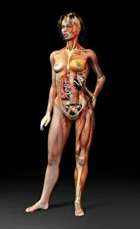
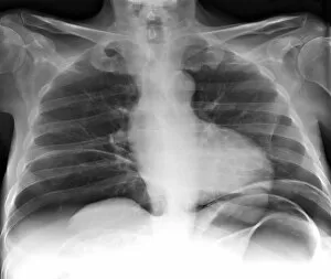
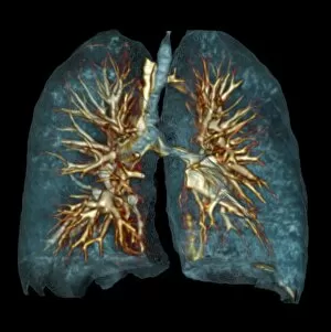
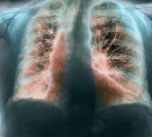
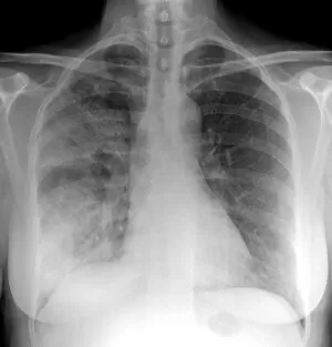
"The Chest: A Canvas of Stories and Secrets" Tattooed masterpiece by Mr. Sutherland Macdonald of Jermyn S: Delicate ink strokes intertwine, revealing a tale etched upon the chest. Tattooed Victorian lady, 1897: Her chest adorned with artistry, a symbol of rebellion against societal norms. Heart: Nestled within the chest, it beats tirelessly, fueling our passions and emotions. Pneumothorax treatment, X-ray: Beneath the surface lies healing; an X-ray unveils the intricate process of mending a wounded chest. Athletic young man in shorts with ball lying on a stone floor: The pounding heart echoes through his chest as he prepares for victory on the court. Maurice Toussaint poster advertising colonial recruitment from World War II, 1938: The call to duty resonates deep within every patriot's chest as they answer their nation's plea. Frank de Burgh, tattooed man, 1897: His body tells stories untold; his vibrant tattoos breathe life into his bare chest. Cardiovascular system historical artwork: An artistic portrayal captures the complexity and beauty that resides within our chests - a network that sustains us all. Vitruvian man with flare in chest: Leonardo da Vinci's masterpiece reveals not only physical proportions but also ignites curiosity about what lies beneath one's own ribcage. Neck anatomy 19th Century artwork: Intricately detailed illustrations unravel the mysteries hidden beneath our throats - connecting voice to soul through this vital passage near the chest. Tuberculosis X-ray : Shadows dance upon fragile lungs captured by an X-ray machine; tuberculosis leaves its mark on countless chests throughout history Ox-pulled wagon - Lipton Tea Company .