
Chloroplasts Collection
Chloroplasts: The Artistry of Nature's Green Machines Cell types come alive through the intricate artwork of chloroplasts


All Professionally Made to Order for Quick Shipping
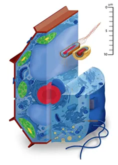
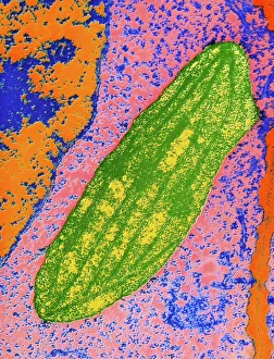
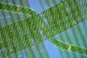
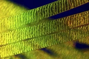
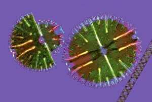
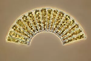
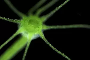
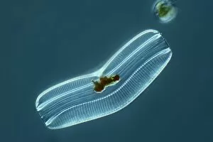
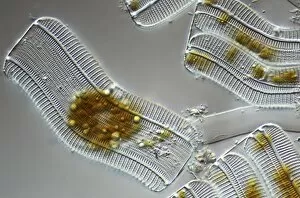
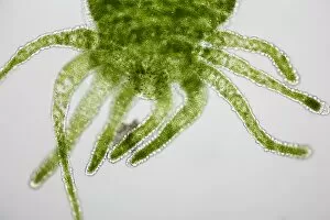
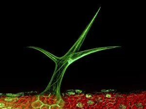
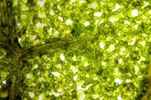
Chloroplasts: The Artistry of Nature's Green Machines Cell types come alive through the intricate artwork of chloroplasts. These tiny, green powerhouses can be found in various plant cells, each contributing to the beauty and functionality of nature's creations. In the cell of a vibrant pea plant, chloroplasts dance with sunlight, capturing its energy for photosynthesis. Their presence gives life to every leaf and fuels growth throughout the plant kingdom. Take a closer look at a tomato leaf under a light micrograph, and you'll witness an enchanting display. Chloroplasts stand out like emerald gems against a backdrop of cellular intricacies, showcasing their importance in converting light into chemical energy. But it doesn't stop there – diatoms reveal their own mesmerizing patterns when viewed under another light micrograph. These microscopic algae showcase chloroplasts that resemble delicate lacework or ornate stained glass windows. Each diatom is adorned with these green organelles that enable them to thrive in aquatic environments. Spirogyra algae also join this artistic spectacle with their spiral-shaped cells containing numerous chloroplasts. Light micrographs capture these organisms as if they were brushstrokes on canvas – vibrant greens intertwining harmoniously within each spirogyra filament. The collaboration between desmids and spirogyra creates yet another masterpiece under the lens of a light micrograph. Here we see an exquisite tapestry where chloroplast-filled cells form intricate patterns reminiscent of Celtic knots or mandalas – proof that even at microscopic levels, nature never ceases to amaze us. Diatoms make another appearance on this visual journey; their unique shapes are accentuated by countless chloroplasts scattered across their silica shells. Like miniature forests captured within glass walls, these diatoms demonstrate how artistry exists even in seemingly simple organisms. Green hydra showcases its elegance as well when observed through a microscope lens. Its translucent body reveals chloroplasts, which provide energy for this remarkable creature.