
Chromatids Collection
Chromatids, the fascinating structures that play a crucial role in cell division and genetic inheritance


All Professionally Made to Order for Quick Shipping
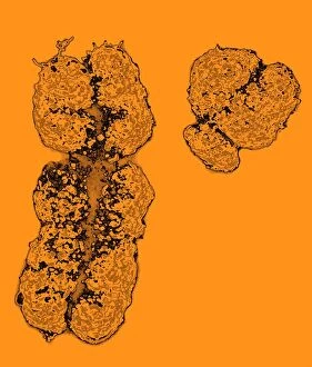
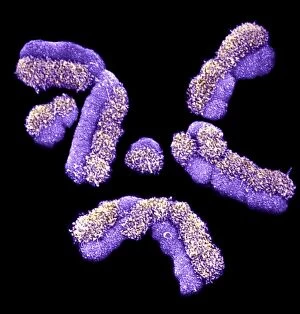
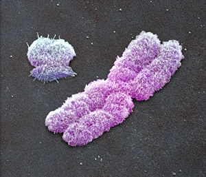
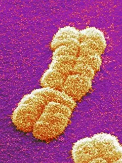
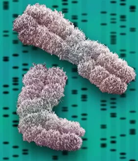
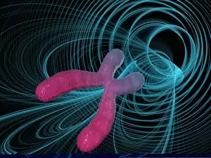
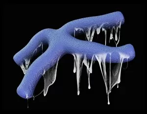
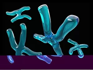
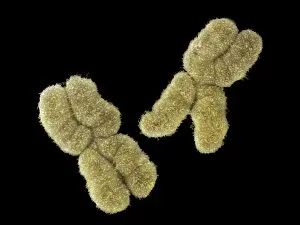
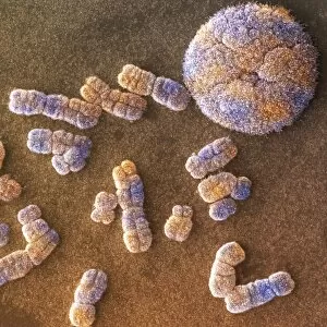
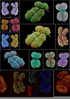
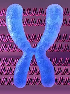
Chromatids, the fascinating structures that play a crucial role in cell division and genetic inheritance. These microscopic threads are responsible for carrying our DNA, including the X and Y chromosomes that determine our biological sex. In mitosis they can be observed under a light micrograph as they align and separate during cell division. This process ensures that each new cell receives an identical set of chromosomes. When it comes to male chromosomes, a full set can be visualized through scanning electron microscopy (SEM). The distinct X and Y chromosomes stand out among others, showcasing their unique roles in determining gender. Dividing cells showcase the intricate dance as they split apart. Each chromosome is duplicated into two sister chromatids connected at the centromere until they eventually segregate into separate cells. Under SEM imaging, human chromosomes appear like delicate strands intricately woven together. The detailed visuals provide insights into their structure and organization within our cells. The captivating beauty of these chromosomal arrangements is evident in SEM images capturing human chromosomes. Their complex patterns highlight the incredible complexity underlying our genetic makeup. As we delve deeper into understanding genetics, studying individual human chromosomes becomes essential. SEM allows us to explore their morphology with remarkable precision, revealing invaluable information about inherited traits and potential health conditions. Chromosomes hold the key to unlocking countless mysteries surrounding life's blueprint. Through SEM imagery, we gain glimpses into their mesmerizing world – a realm where genes intertwine to shape who we are as individuals. So next time you marvel at those microscopic wonders called chromatids or gaze upon stunning images of human chromosomes captured by SEM technology, remember how these tiny entities hold immense power in shaping life itself.