Fraud Blocker Skip to main content

Cilia Collection

Cilia: The Tiny Powerhouses of the Microscopic World Step into the fascinating realm of cilia

Background imageCilia Collection: Brain surface, SEM

Brain surface, SEM
Brain surface. Coloured scanning electron micrograph (SEM) of the surface of a brain ventricle. Ventricles are cavities within the brain that are continuous with the central canal of the spinal cord

Background imageCilia Collection: Light Micrograph (LM): Protozoans: Kidney shaped ciliate surrounded by Euglena sp

Light Micrograph (LM): Protozoans: Kidney shaped ciliate surrounded by Euglena sp
LRDS-316 Light Micrograph (LM): Protozoans: Kidney shaped ciliate surrounded by Euglena sp. Magnification x 900 (when printed A4, 29.7 cm wide) Tillina sp

Background imageCilia Collection: Trachea lining, SEM

Trachea lining, SEM
Trachea lining, coloured scanning electron micrograph (SEM). The tracheal lining contains mucus-secreting goblet cells and epithelial cells bearing hair-like cilia (blue)

Background imageCilia Collection: Spider lily flower stamen, SEM

Spider lily flower stamen, SEM
Spider lily flower stamen. Coloured scanning electron micrograph (SEM) of a cilia (hair-like structures) from a spider lily flower (Hymenocallis sp.) stamen

Background imageCilia Collection: Picture No. 11014599

Picture No. 11014599
Freshwater Ciliates common in pools and ponds consisting of a single cell and feed by ingesting other tiny organisms such as bacteria and other protozoa Date:

Background imageCilia Collection: Inner ear hairs, SEM

Inner ear hairs, SEM
Inner ear hairs. Coloured scanning electron micrograph (SEM) of stereocilia, sensory hair-like projections, from hair cells of the cochlea of the inner ear

Background imageCilia Collection: Crown Jellyfish, Cephea cephea, Hawaii, USA

Crown Jellyfish, Cephea cephea, Hawaii, USA
The Crown jellyfish (Cephea cephea) is also known as the cauliflower jellyfish, Hawaii. This species lives in open ocean with a lifespan of three to six months

Background imageCilia Collection: Paramecium sp. protozoan, SEM

Paramecium sp. protozoan, SEM
Paramecium protozoan. Coloured scanning electron micrograph (SEM) of a Paramecium sp. protozoan. This single-celled organism is aquatic, living in freshwater habitats

Background imageCilia Collection: Nasal lining, SEM

Nasal lining, SEM
Nasal lining. Coloured scanning electron micrograph (SEM) of the olfactory epithelium that lines the nasal cavity, showing olfactory cells (red) surrounded by numerous cilia (hair-like projections)

Background imageCilia Collection: Trachea lining, SEM C015 / 9936

Trachea lining, SEM C015 / 9936
Trachea lining. Coloured scanning electron micrograph (SEM) of a section through the lining of the trachea (wind pipe). The trachea links the larynx (voice box) to the lungs

Background imageCilia Collection: Trachea lining, SEM C013 / 7126

Trachea lining, SEM C013 / 7126
Trachea lining. Coloured scanning electron micrograph (SEM) of the lining of a trachea (wind pipe), showing the cilia (hair-like) and goblet cells (orange). The trachea links the larynx to the lungs

Background imageCilia Collection: Trachea lining, SEM C013 / 7122

Trachea lining, SEM C013 / 7122
Trachea lining. Coloured scanning electron micrograph (SEM) of a section through the wall of a trachea (wind pipe). The trachea links the larynx to the lungs

Background imageCilia Collection: Paramecium bursaria protozoan, micrograph

Paramecium bursaria protozoan, micrograph
Paramecium bursaria protozoan, light micrograph. This ciliate protozoan inhabits freshwater, where it feeds mainly on bacteria

Background imageCilia Collection: Allergens in trachea

Allergens in trachea. Coloured scanning electron micrograph (SEM) of the surface of the trachea (windpipe) with a number of pollen grains (yellow) attached

Background imageCilia Collection: Trachea, SEM

Trachea, SEM
Trachea. Coloured scanning electron micrograph (SEM) of the lining of the trachea (windpipe). The trachea links the larynx (voice box) to the lungs

Background imageCilia Collection: Paramecium protozoa, light micrograph

Paramecium protozoa, light micrograph
Paramecium bursaria protozoa, light micrograph. These ciliate protozoa inhabit freshwater, where they feed mainly on bacteria

Background imageCilia Collection: Trichodina parasite, light micrograph

Trichodina parasite, light micrograph
Trichodina parasite. Differential interference contrast micrograph of Trichodina pediculus. This ciliate parasite lives on the skin, gills and mucous membranes of many aquatic animals

Background imageCilia Collection: Bacteria on lung hairs

Bacteria on lung hairs
Bacteria in a lung. Computer artwork of bacteria (yellow) on cilia (hair-like structures) in a lung. Bacterial infection of the lungs can cause potentially fatal pneumonia

Background imageCilia Collection: Illustration of Trochophore, a transparent marine larva showing internal organs

Illustration of Trochophore, a transparent marine larva showing internal organs and abundance of external cilia

Background imageCilia Collection: Illustration of Blue Mussel (Mytilus edulis), showing oval opening and fringed opening

Illustration of Blue Mussel (Mytilus edulis), showing oval opening and fringed opening, and barnacles growing on shell

Background imageCilia Collection: Picture No. 11675491

Picture No. 11675491
Light Micrograph: Ciliate. Date:

Background imageCilia Collection: Picture No. 11675490

Picture No. 11675490
Light Micrograph (LM): Elongate Ciliate and Paramecium bursaria. Date:

Background imageCilia Collection: Trachea lining, SEM C016 / 9756

Trachea lining, SEM C016 / 9756
Trachea lining. Coloured scanning electron micrograph (SEM) of the lining of the trachea (wind pipe). The trachea links the larynx to the lungs

Background imageCilia Collection: Protozoan sexual reproduction, SEM Z105 / 0240

Protozoan sexual reproduction, SEM Z105 / 0240
Protozoan sexual reproduction. Coloured scanning electron micrograph (SEM) of two Paramecium caudatum protozoa reproducing sexually by a method called conjugation

Background imageCilia Collection: Paramecium protozoa, SEM Z105 / 0239

Paramecium protozoa, SEM Z105 / 0239
Paramecium protozoa. Coloured scanning electron micrograph (SEM) of two Paramecium caudatum protozoa on filaments (green) of Oscillatoria sp. blue-green algae. P

Background imageCilia Collection: Ampullary cupula, artwork

Ampullary cupula, artwork
Ampullary cupula. Computer artwork showing how the ampullary cupula in the human vestibular system of the inner ear senses head rotation (top) and forward motion (bottom)

Background imageCilia Collection: Holosticha ciliate protozoan, SEM

Holosticha ciliate protozoan, SEM
Holosticha ciliate protozoan. Coloured scanning electron micrograph (SEM) of a Holosticha sp. ciliate protozoan ingesting diatoms (blue). Holosticha is a tiny single-celled aquatic organism

Background imageCilia Collection: Euplotes protozoa, SEM C016 / 9040

Euplotes protozoa, SEM C016 / 9040
Euplotes protozoa. Coloured scanning electron micrograph (SEM) showing the upperside (bottom) and underside (top) of two Euplotes sp. hypotrich ciliate protozoa

Background imageCilia Collection: Climacostomum protozoan, SEM C016 / 9063

Climacostomum protozoan, SEM C016 / 9063
Climacostomum protozoan. Coloured scanning electron micrograph (SEM) of a Climacostomum sp. unicellular ciliate protozoan, showing the cells large oral apparatus (round, centre)

Background imageCilia Collection: Climacostomum protozoan, SEM C016 / 9121

Climacostomum protozoan, SEM C016 / 9121
Climacostomum protozoan. Coloured scanning electron micrograph (SEM) of a Climacostomum sp. unicellular ciliate protozoan, showing the cells large oral apparatus (round, centre)

Background imageCilia Collection: Climacostomum protozoan, SEM C016 / 9061

Climacostomum protozoan, SEM C016 / 9061
Climacostomum protozoan. Coloured scanning electron micrograph (SEM) of a Climacostomum sp. unicellular ciliate protozoan, showing the cells large oral apparatus (round, upper right)

Background imageCilia Collection: Climacostomum protozoan, SEM C016 / 9120

Climacostomum protozoan, SEM C016 / 9120
Climacostomum protozoan. Coloured scanning electron micrograph (SEM) of a Climacostomum sp. unicellular ciliate protozoan, showing the cells large oral apparatus (round, centre)

Background imageCilia Collection: Climacostomum protozoan, SEM C016 / 9065

Climacostomum protozoan, SEM C016 / 9065
Climacostomum protozoan. Coloured scanning electron micrograph (SEM) of a Climacostomum sp. unicellular ciliate protozoan, showing the cells large oral apparatus (round, upper right)

Background imageCilia Collection: Climacostomum protozoan, SEM C016 / 9064

Climacostomum protozoan, SEM C016 / 9064
Climacostomum protozoan. Coloured scanning electron micrograph (SEM) of a Climacostomum sp. unicellular ciliate protozoan, showing the cells large oral apparatus (round, centre)

Background imageCilia Collection: Climacostomum protozoan, SEM C016 / 9122

Climacostomum protozoan, SEM C016 / 9122
Climacostomum protozoan. Coloured scanning electron micrograph (SEM) of a Climacostomum sp. unicellular ciliate protozoan, showing the cells large oral apparatus (round, centre)

Background imageCilia Collection: Euplotes protozoa, SEM C016 / 9041

Euplotes protozoa, SEM C016 / 9041
Euplotes protozoa. Coloured scanning electron micrograph (SEM) showing the upperside (bottom) and underside (top) of two Euplotes sp. hypotrich ciliate protozoa

Background imageCilia Collection: Climacostomum protozoan, SEM C016 / 9062

Climacostomum protozoan, SEM C016 / 9062
Climacostomum protozoan. Coloured scanning electron micrograph (SEM) of a Climacostomum sp. unicellular ciliate protozoan, showing the cells large oral apparatus (round, upper right)

Background imageCilia Collection: Ciliate protozoan ingesting algae

Ciliate protozoan ingesting algae. Darkfield illuminated light micrograph of a ciliate protozoan ingesting green algae (green)

Background imageCilia Collection: Tetrahymena protozoan, SEM

Tetrahymena protozoan, SEM
Tetrahymena protozoan. Coloured scanning electron micrograph (SEM) of a Tetrahymena sp. ciliate protozoan. Tetrahymena are free-living freshwater unicellular organisms found in ponds

Background imageCilia Collection: Smell receptor, TEM

Smell receptor, TEM
Smell receptor. Transmission electron micrograph (TEM) of a section through the olfactory epithelium that lines the nasal cavity, showing an olfactory cell (smell receptor)

Background imageCilia Collection: Ciliate protozoan, light micrograph

Ciliate protozoan, light micrograph
Ciliate protozoan. Darkfield illuminated light micrograph of a ciliate protozoan, showing green algae (green) that it has ingested

Background imageCilia Collection: Trachea lining, SEM C015 / 9939

Trachea lining, SEM C015 / 9939
Trachea lining. Coloured scanning electron micrograph (SEM) of a section through the lining of the trachea (wind pipe). The trachea links the larynx (voice box) to the lungs

Background imageCilia Collection: Trachea lining, SEM C015 / 9610

Trachea lining, SEM C015 / 9610
Trachea lining. Coloured scanning electron micrograph (SEM) of the lining of the trachea (wind pipe). The trachea links the larynx to the lungs

Background imageCilia Collection: Trachea lining, SEM C015 / 9609

Trachea lining, SEM C015 / 9609
Trachea lining, SEM

Background imageCilia Collection: Sperm cells, artwork C018 / 7004

Sperm cells, artwork C018 / 7004
Sperm cells. Computer artwork of numerous sperm cells (spermatozoa) swimming through the cilia (hair-like) that line the uterus (womb)

Background imageCilia Collection: Trachea lining, SEM C016 / 9755

Trachea lining, SEM C016 / 9755
Trachea lining. Coloured scanning electron micrograph (SEM) of the lining of the trachea (wind pipe). The trachea links the larynx to the lungs


All Professionally Made to Order for Quick Shipping

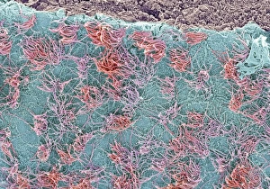
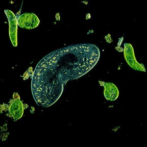
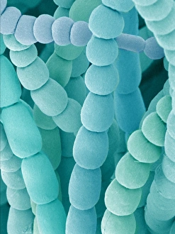
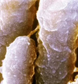
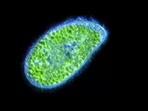
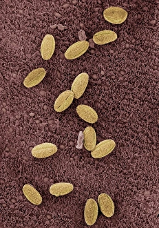
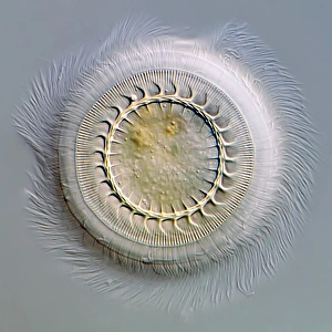
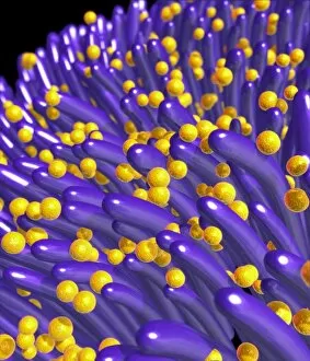
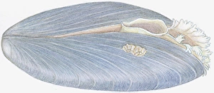
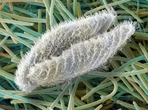
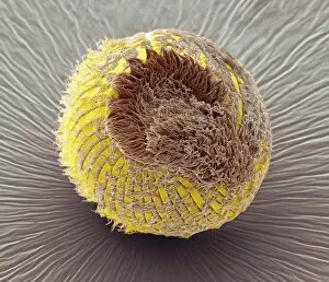
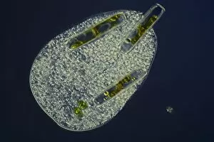
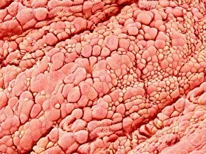
Cilia: The Tiny Powerhouses of the Microscopic World Step into the fascinating realm of cilia, where these tiny hair-like structures play a crucial role in various organisms. Captured under the lens of a light microscope (LM) at an impressive magnification of x900, we witness their intricate beauty and functionality. In one captivating image, we observe kidney-shaped ciliates surrounded by Euglena sp. , showcasing the diversity within protozoans. These delicate creatures rely on their cilia for locomotion and feeding, highlighting their vital importance in this microscopic ecosystem. Moving to another scene, an SEM image reveals the brain surface adorned with countless cilia. These microscopic hairs serve as sensory receptors, allowing neurons to communicate and process information efficiently. Venturing further down our exploration path, we encounter trachea linings captured through scanning electron microscopy (SEM). Here, cilia take center stage once again as they tirelessly beat in unison to clear mucus and foreign particles from our respiratory system—a remarkable defense mechanism that keeps us breathing freely. Nature's artistic touch is revealed in stunning detail when examining spider lily flower stamens using SEM. Amongst vibrant petals lies a hidden world of intricately arranged cilia that aid in pollen capture and dispersal—an essential step towards plant reproduction. Delving deeper into the human body's wonders, SEM exposes brain linings where specialized cells extend numerous cilia. These fine projections facilitate cerebrospinal fluid circulation while also contributing to sensory perception—a testament to nature's ingenious design. The fallopian tube cells come into focus next; SEM captures their elegant structure embellished with clusters of meticulously aligned cilia. This arrangement ensures smooth transportation of eggs towards fertilization sites—nature's way of nurturing life itself. Picture No. 11014599 unveils yet another marvel—the inner ear hairs observed through SEM. Ciliated cells here enable hearing by converting sound vibrations into electrical signals, allowing us to appreciate the symphony of life.