
Ciliate Collection
Ciliates, a fascinating group of unicellular eukaryotic organisms, are the stars of these captivating micrographs and illustrations


All Professionally Made to Order for Quick Shipping
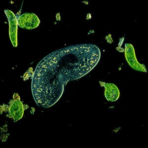
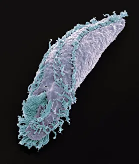
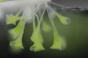
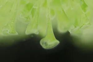
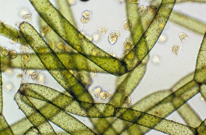
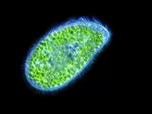
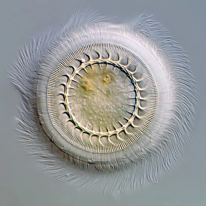
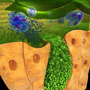
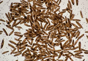
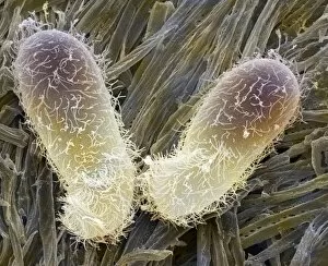
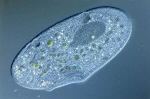
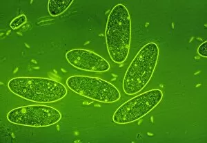
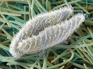
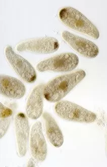
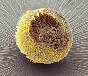
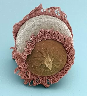
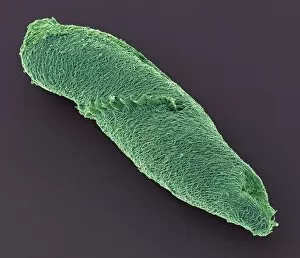
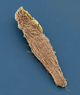
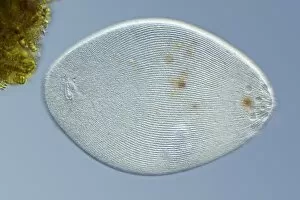
Ciliates, a fascinating group of unicellular eukaryotic organisms, are the stars of these captivating micrographs and illustrations. In one LM image, we can observe a kidney-shaped ciliate surrounded by Euglena sp. , magnified 900 times to reveal intricate details that would otherwise go unnoticed. Another SEM image showcases Oxytricha ciliate protozoan, providing us with a closer look at its unique structure. Moving on to the trumpet animalcules (Stentor polymorphus), we discover their symbiotic relationship with algal partners called Chlorella. These microscopic creatures can be found in Europe and form an intriguing alliance that benefits both parties involved. Vorticella ciliates take center stage in another LM picture as they gracefully attach themselves to a green alga. Their elegant appearance is truly mesmerizing under the lens of a microscope. Paramecium sp. , another type of protozoan, also makes an appearance in this collection through both SEM and micrograph images showcasing their distinct features. Not only do these images showcase the beauty and diversity of ciliates but they also shed light on their interactions within ecosystems. For instance, Trichodina parasite is captured in a light micrograph highlighting its parasitic nature. Lastly, an illustration provides us with a digital cross-section view of a ciliate cell residing in the nasal cavity alongside rhinovirus particles and antibodies—an artistic representation that merges science and creativity seamlessly. These glimpses into the world of ciliates remind us how much there still is to explore within our own microscopic backyard—a realm teeming with life waiting for curious minds to uncover its secrets.