
Cilium Collection
"Cilia: The Tiny Powerhouses of the Microscopic World" Cilia, those hair-like structures found in various parts of our body and beyond, are truly remarkable


All Professionally Made to Order for Quick Shipping
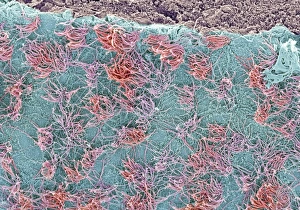
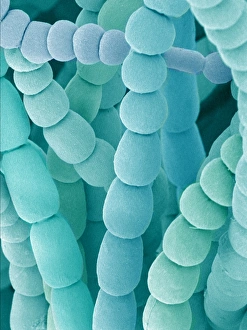
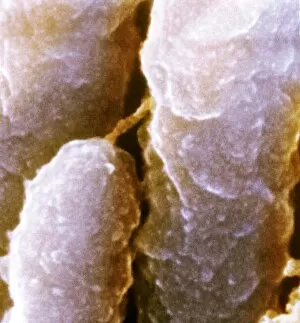
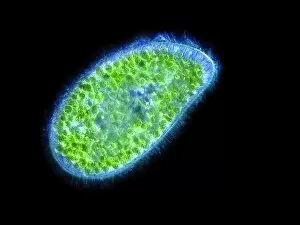
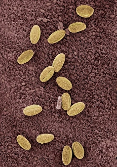
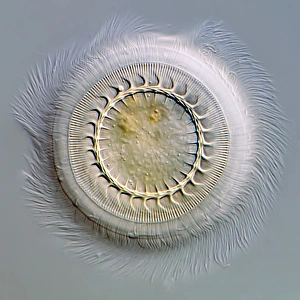
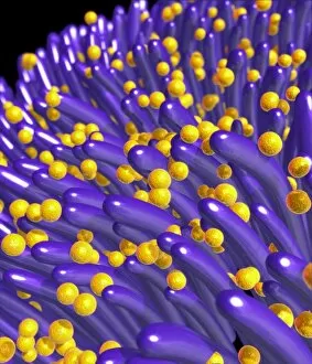
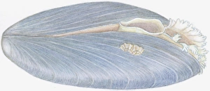
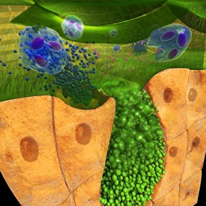
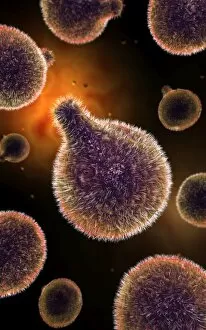
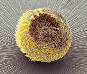
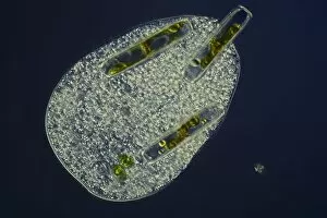
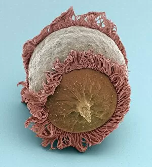
"Cilia: The Tiny Powerhouses of the Microscopic World" Cilia, those hair-like structures found in various parts of our body and beyond, are truly remarkable. From the brain surface to the trachea lining, from spider lily flower stamen to fallopian tube cells, these tiny appendages play vital roles in different organisms. Underneath a scanning electron microscope (SEM), we can witness the intricate beauty of cilia. In the brain lining, they form a dense carpet-like layer that helps facilitate communication between neurons and ensures efficient functioning of our nervous system. Moving down to the trachea lining, SEM reveals an astonishing sight. Cilia here act as nature's broomsticks, constantly beating in unison to sweep away mucus and foreign particles that enter our respiratory system. They serve as guardians protecting us from harmful invaders. Even plants have their own version of cilia. Spider lily flower stamen showcases delicate filaments with ciliary structures under SEM examination. These specialized hairs aid in pollination by capturing pollen grains and facilitating their transfer for reproduction. Intriguingly, fallopian tubes also possess ciliated cells that assist in guiding eggs towards fertilization sites within a woman's reproductive system. Under SEM scrutiny, these microscopic hairs appear like miniature oars propelling life forward on its journey towards conception. Venturing into another realm altogether – inner ear hairs – we discover yet another crucial function performed by cilia. These sensory receptors help convert sound vibrations into electrical signals that allow us to perceive sounds and maintain balance. The world beneath an SEM doesn't limit itself solely to humans; it extends even further into microorganisms such as Paramecium sp. , where countless tiny cilia cover their entire bodies. These protozoans use synchronized movements of their numerous cilia for locomotion through water environments. Returning back to human anatomy once more - nasal linings reveal complex arrangements of cilia that serve as the first line of defense against airborne pathogens.