Fraud Blocker Skip to main content

Compound Eye Collection (page 3)

The compound eye, a marvel of nature's engineering, is a fascinating feature found in various insects

Background imageCompound Eye Collection: Gall midge eye, SEM C016 / 9394

Gall midge eye, SEM C016 / 9394
Gall midge eye. Coloured scanning electron micrograph (SEM) of the compound eye of a gall midge (family Cecidomyiidae), showing the individual lenses (ommatidia)

Background imageCompound Eye Collection: Owl butterfly head, SEM C016 / 9432

Owl butterfly head, SEM C016 / 9432
Owl butterfly head. Coloured scanning electron micrograph (SEM) of the head of an owl butterfly (Caligo sp.), showing its two large compound eyes (blue) and antennae (upper left and right)

Background imageCompound Eye Collection: Fly eye, SEM C016 / 9392

Fly eye, SEM C016 / 9392
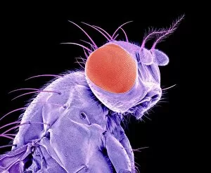
Fly eye. Coloured scanning electron micrograph (SEM) of the compound eye (red) of a fly (order Diptera), showing the individual lenses (ommatidia, right) and hairs (blue)

Background imageCompound Eye Collection: Common blue damselfly eyes, SEM C016 / 9383

Common blue damselfly eyes, SEM C016 / 9383
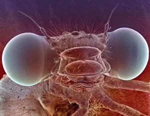
Common blue damselfly eyes, SEM. Coloured scanning electron micrograph (SEM) of the head of a common blue damselfly (Enallagma cyathigerum), showing its large compound eyes

Background imageCompound Eye Collection: Firefly head, SEM C016 / 9413

Firefly head, SEM C016 / 9413
Firefly head. Coloured scanning electron micrograph (SEM) of the head of a firefly (family Lampyridae), showing its compound eyes (dark green) and antennae (upper left and right)

Background imageCompound Eye Collection: Grasshopper head

Grasshopper head. Close-up of the head of a grasshopper (order Orthoptera), showing one of its compound eyes (black). Magnification: x33, when printed 10 centimetres wide

Background imageCompound Eye Collection: Common darter dragonfly

Common darter dragonfly. Close-up of a common darter (Sympetrum striolatum) dragonfly showing its large compound eyes (top). Photographed in September

Background imageCompound Eye Collection: Insect eye anatomy, artwork

Insect eye anatomy, artwork
Insect eye anatomy. Computer artwork showing the structure of a compound eye from an insect. Compound eyes are made up of individual lenses called ommatidia (hexagonal, blue, and far right)

Background imageCompound Eye Collection: Fly, SEM C015 / 8077

Fly, SEM C015 / 8077
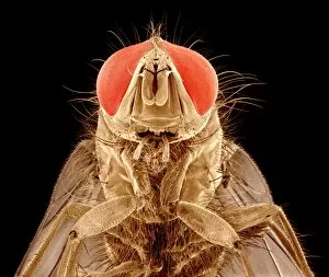
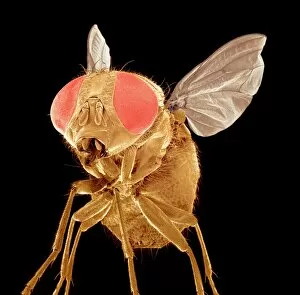
Fly. Coloured scanning electron micrograph (SEM) of a fly (order Diptera). The head is dominated by two large compound eyes (red)

Background imageCompound Eye Collection: Moth head, SEM C015 / 8073

Moth head, SEM C015 / 8073
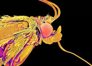
Moth head, coloured scanning electron micrograph (SEM). The head is dominated by the large compound eyes (one seen, pink). Two antennae are mounted on the top of the head

Background imageCompound Eye Collection: Moth head, SEM C015 / 8074

Moth head, SEM C015 / 8074
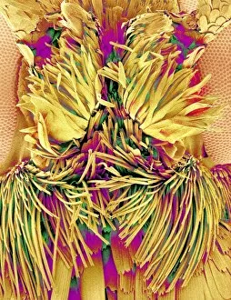
Moth head. coloured scanning electron micrograph (SEM) of the area of the head between the moths compound eyes (pink, far right and far left). Two antennae are mounted at upper centre

Background imageCompound Eye Collection: Fly, SEM C015 / 9945

Fly, SEM C015 / 9945
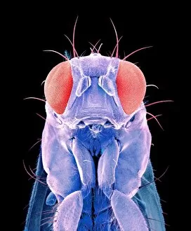
Fly. Coloured scanning electron micrograph (SEM) of a fly (order Diptera). The head is dominated by two large compound eyes (red)

Background imageCompound Eye Collection: Moth head, SEM C015 / 8071

Moth head, SEM C015 / 8071
Moth head, coloured scanning electron micrograph (SEM). The head is dominated by the large compound eyes (one seen, pink). Two antennae are mounted on the top of the head

Background imageCompound Eye Collection: Fly, SEM C015 / 8080

Fly, SEM C015 / 8080
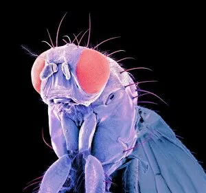
Fly. Coloured scanning electron micrograph (SEM) of a fly (order Diptera). The head is dominated by two large compound eyes (red)

Background imageCompound Eye Collection: Fly, SEM C015 / 8082

Fly, SEM C015 / 8082
Fly. Coloured scanning electron micrograph (SEM) of a fly (order Diptera). The head is dominated by two large compound eyes (red)

Background imageCompound Eye Collection: African mantis C016 / 2288

African mantis C016 / 2288
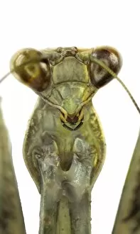
African mantis. Close-up of the head of a giant African mantis (Sphodromantis viridis), showing one of its large compound eyes (centre)

Background imageCompound Eye Collection: Moth head, SEM C015 / 8072

Moth head, SEM C015 / 8072
Moth head, coloured scanning electron micrograph (SEM). The head is dominated by the large compound eyes (one seen, pink). Two antennae are mounted on the top of the head

Background imageCompound Eye Collection: Fly, SEM C015 / 8081

Fly, SEM C015 / 8081
Fly. Coloured scanning electron micrograph (SEM) of a fly (order Diptera). The head is dominated by two large compound eyes (one seen, red)

Background imageCompound Eye Collection: African mantis C016 / 2283

African mantis C016 / 2283
African mantis (Sphodromantis viridis). This species of praying mantis is kept worldwide as a pet but is native to West Africa, south of the Sahara Desert

Background imageCompound Eye Collection: Fly, SEM C015 / 8078

Fly, SEM C015 / 8078
Fly. Coloured scanning electron micrograph (SEM) of a fly (order Diptera). The head is dominated by two large compound eyes (red)

Background imageCompound Eye Collection: African mantis C016 / 2285

African mantis C016 / 2285
African mantis. Close-up of the head of a giant African mantis (Sphodromantis viridis), showing its large compound eyes (upper left and right)

Background imageCompound Eye Collection: Fly, SEM C015 / 8079

Fly, SEM C015 / 8079
Fly. Coloured scanning electron micrograph (SEM) of a fly (order Diptera). The head is dominated by two large compound eyes (red)

Background imageCompound Eye Collection: Fly, SEM C015 / 9944

Fly, SEM C015 / 9944
Fly. Coloured scanning electron micrograph (SEM) of a fly (order Diptera). The head is dominated by two large compound eyes (red)

Background imageCompound Eye Collection: Fly, SEM C015 / 8083

Fly, SEM C015 / 8083
Fly. Coloured scanning electron micrograph (SEM) of a fly (order Diptera). The head is dominated by two large compound eyes (red)

Background imageCompound Eye Collection: Insect compound eye, SEM C018 / 0554

Insect compound eye, SEM C018 / 0554
Insect compound eye, coloured scanning electron micrograph (SEM). The units in a compound eye, the ommatidia, each contain a cornea and photoreceptor cells

Background imageCompound Eye Collection: Electron microscopy water artefact, SEM C018 / 0557

Electron microscopy water artefact, SEM C018 / 0557
Electron microscopy water artefact, coloured scanning electron micrograph (SEM). This is the surface of a compound eye from a bee

Background imageCompound Eye Collection: Ant head, SEM C018 / 0547

Ant head, SEM C018 / 0547
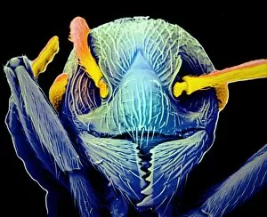
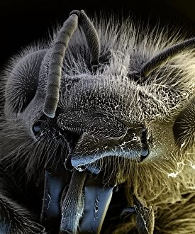
Ant head, coloured scanning electron micrograph (SEM). The antennae (sensory structures) are the stalk-like structures (orange) attached to the head above the mouthparts (bottom)

Background imageCompound Eye Collection: Insect compound eye, SEM C018 / 0553

Insect compound eye, SEM C018 / 0553
Insect compound eye, coloured scanning electron micrograph (SEM). The units in a compound eye, the ommatidia, each contain a cornea and photoreceptor cells

Background imageCompound Eye Collection: Ant head, SEM C018 / 0548

Ant head, SEM C018 / 0548
Ant head, coloured scanning electron micrograph (SEM). The antennae (sensory structures) are the stalk-like structures (blue) attached to the head above the mouthparts (bottom)

Background imageCompound Eye Collection: Fly compound eye lenses, SEM C018 / 0556

Fly compound eye lenses, SEM C018 / 0556
Fly compound eye lenses, coloured scanning electron micrograph (SEM). A compound eye consists of many rounded lenses known as ommatidia

Background imageCompound Eye Collection: Lesser stag beetle female, SEM C018 / 0317

Lesser stag beetle female, SEM C018 / 0317
Lesser stag beetle (Dorcus parallelipipedus) female, coloured scanning electron micrograph (SEM). Visible here on its head are its mouthparts, antennae and one of its eyes

Background imageCompound Eye Collection: Fruit fly compound eye lenses, SEM C018 / 0555

Fruit fly compound eye lenses, SEM C018 / 0555
Fruit fly compound eye lenses, coloured scanning electron micrograph (SEM). A compound eye consists of many rounded lenses known as ommatidia

Background imageCompound Eye Collection: Fruit fly head C018 / 5815

Fruit fly head C018 / 5815
Fruit fly (Tephritis postica) head. Each compound eye (green) is made up of numerous simple eyes called ommatidia. Each ommatidium sends a signal to the flys brain

Background imageCompound Eye Collection: Grasshopper C018 / 4807

Grasshopper C018 / 4807
Grasshopper (Melanoplus sanguinipes), side view photograph. This specimen was found in Maryland, USA. Photographed by the USGS Bee Inventory and Monitoring Lab

Background imageCompound Eye Collection: Spider wasp C018 / 4318

Spider wasp C018 / 4318
Spider wasp (Pompilidae), close-up photo. This specimen (species unknown) was found in the San Bernarndino, California, USA. Spider wasps are solitary and hunt spiders

Background imageCompound Eye Collection: Cuckoo wasp C018 / 4317

Cuckoo wasp C018 / 4317
Cuckoo wasp, close-up photo. This specimen (species unknown) was found in Timpanogos Cave National Monument, Utah, USA. Cuckoo wasps (Chrysididae family)

Background imageCompound Eye Collection: Pygmy grasshopper C018 / 4809

Pygmy grasshopper C018 / 4809
Pygmy grasshopper (Tettigidea lateralis), close-up photograph. This specimen was found in Maryland, USA. Photographed by the USGS Bee Inventory and Monitoring Lab

Background imageCompound Eye Collection: Male bee C018 / 3571

Male bee C018 / 3571
Male bee. Close-up photograph of a male bee head (Anthophora affabilis). This specimen is native to the US and was found in Badlands National Park, South Dakota

Background imageCompound Eye Collection: Indian hemp beetle C018 / 4686

Indian hemp beetle C018 / 4686
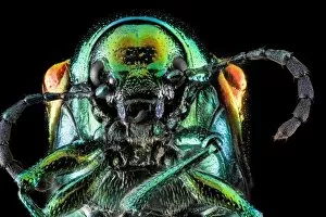
Indian hemp beetle (Chrysochus auratus), close-up photograph. This specimen was found in Maryland, USA. It has an iridescent blue-green coloured body

Background imageCompound Eye Collection: Apple bark borer moth C018 / 4806

Apple bark borer moth C018 / 4806
Apple bark borer moth (Synanthedon pyri) close-up photograph of the head and thorax. The moth is covered with iridescent coloured scales. This specimen was found in Maryland, USA

Background imageCompound Eye Collection: Female bee head C018 / 3570

Female bee head C018 / 3570
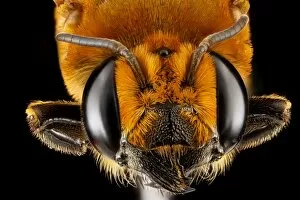
Female bee head. Close-up photograph of a female bee head (Megachile lanata). This specimen was found in Cuba. The species was originally from Africa and has since spread to the Caribbean

Background imageCompound Eye Collection: Female blue mud dauber wasp C018 / 4685

Female blue mud dauber wasp C018 / 4685
Female blue mud dauber wasp (Chalybion californicum), side view. This species is native to North America. It is coloured metallic blue and preys on black widow spiders

Background imageCompound Eye Collection: Damselfly eye

Damselfly eye. Close-up of one of the compound eyes of a damselfly (order Odonata), showing the individual lenses (ommatidia, hexagonal)

Background imageCompound Eye Collection: Bluebottle

Bluebottle. Close-up of the head of a bluebottle (family Calliphoridae) fly, showing its large compound eyes (centre left and right). Magnification: x34 when printed 10 centimetres wide

Background imageCompound Eye Collection: Fly eye, SEM C014 / 4848

Fly eye, SEM C014 / 4848
Fly eye. Coloured scanning electron micrograph (SEM) showing the lenses (ommatidia, hexagonal) that make up the compound eye of a fly

Background imageCompound Eye Collection: Wasp

Wasp. Close-up of the head of a wasp (order Hymenoptera), showing its large compound eyes (black, left and right). Magnification: x17 when printed 10 centimetres wide

Background imageCompound Eye Collection: Honey bee head, SEM C016 / 8018

Honey bee head, SEM C016 / 8018
Honey bee head. Coloured scanning electron micrograph (SEM) of the head of a honey bee (Apis sp.), showing its large compound eyes (left and right), mouthparts (lower centre)

Background imageCompound Eye Collection: Honey bee head, SEM C016 / 8022

Honey bee head, SEM C016 / 8022
Honey bee head. Coloured scanning electron micrograph (SEM) of the head of a honey bee (Apis sp.), showing its large compound eyes (left and right), mouthparts (lower centre)


All Professionally Made to Order for Quick Shipping

The compound eye, a marvel of nature's engineering, is a fascinating feature found in various insects. Take the fruit fly for instance, as seen through the lens of an SEM Z340 / 0768. Magnified at x300 on an A4 size frame, its compound eye reveals intricate details that captivate our imagination. But it's not just the fruit fly that showcases this incredible structure. The Culex mosquito and red-barbed ant also possess compound eyes that have been meticulously examined under scanning electron micrographs (SEM). These images provide us with glimpses into their world, where vision takes on a whole new dimension. Moving on to the head of a honey bee captured by another SEM image, we witness how these tiny creatures rely heavily on their compound eyes for navigation and finding nectar-rich flowers amidst vast landscapes, and is truly remarkable how such small organisms can possess such complex visual systems. And let's not forget about other species like the hornet mimic hoverfly or even mosquitoes themselves. Their internal anatomy has been revealed through cross-sections and SEM images showcasing their feeding habits - including blood-sucking from human skin. Zooming back in to focus solely on flies, we encounter yet another stunning view of a house fly's compound eye magnified at x40 using SEM technology. This close-up view allows us to appreciate the intricate arrangement of individual lenses called ommatidia which make up this unique visual organ. With beekeeping being an essential practice worldwide, understanding the compound eye becomes crucial in managing beehives effectively. By comprehending how bees perceive their surroundings through these specialized organs, beekeepers can ensure optimal conditions for honey production and colony health. Finally returning to our initial subject - the fruit fly - we delve deeper into its microscopic world with another SEM image (Z340 / 0699).