
Computer Tomography Collection
Computer tomography, also known as CT scan, is a revolutionary medical imaging technique that has transformed the way we diagnose and treat various conditions


All Professionally Made to Order for Quick Shipping
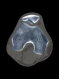
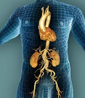
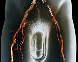
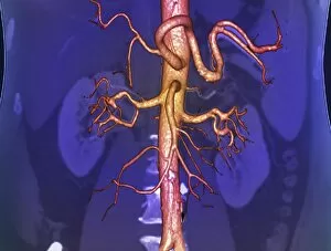
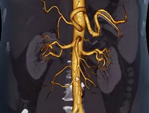
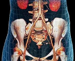
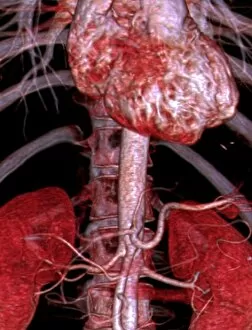
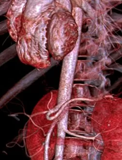
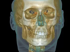
Computer tomography, also known as CT scan, is a revolutionary medical imaging technique that has transformed the way we diagnose and treat various conditions. With its ability to provide detailed cross-sectional images of the body, CT scans have become an indispensable tool in modern medicine. One area where CT scans have proven particularly beneficial is in diagnosing knee diseases. By using a 3D CT scan (F006 / 9140), doctors can accurately visualize the affected area and identify any abnormalities or damage present. This allows for precise diagnosis and targeted treatment plans tailored to each patient's specific needs. Another crucial application of CT scanning is in examining the aorta, the main artery that carries blood from the heart to the rest of the body. Through advanced technology like 3D CT scans (F006 / 9120) and (F006 / 9119), doctors can assess any potential issues such as aneurysms or blockages within this vital blood vessel. Early detection through these non-invasive imaging techniques enables prompt intervention, potentially saving lives. CT scans are not limited to specific areas but encompass various parts of our anatomy. For instance, when studying chest anatomy, 3D CT scans (F006 / 9118), (F006 / 9117), and (F006 / 9116) offer comprehensive insights into lung health and other thoracic structures. These images assist physicians in identifying abnormalities such as tumors or infections promptly. Moreover, computer tomography plays a significant role in evaluating neck bones' normal structure using a detailed 3D CT scan (F006 / 9115). This aids orthopedic specialists in assessing bone density and detecting fractures or degenerative changes effectively. Furthermore, by utilizing high-resolution imaging techniques like narrow arteries' visualization with a 3D CT scan (F006/9113), healthcare professionals can detect arterial stenosis early on—a condition characterized by narrowed blood vessels that can lead to serious complications like heart attacks or strokes.