
Corpus Callosum Collection
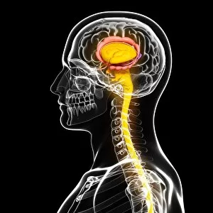
The corpus callosum, a vital structure in the human brain, serves as a bridge connecting the two hemispheres


All Professionally Made to Order for Quick Shipping
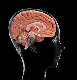
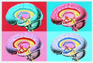
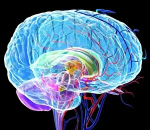
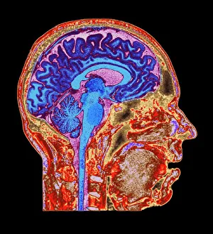
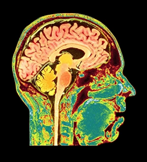
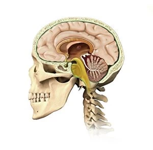
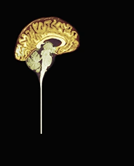
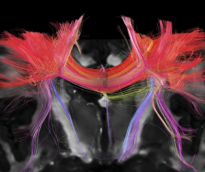
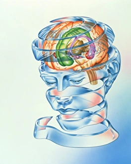
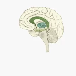
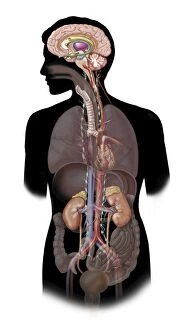
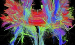
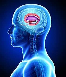
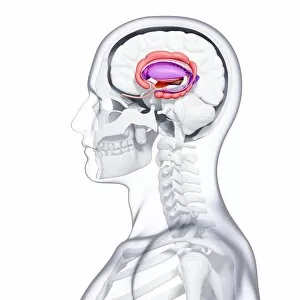
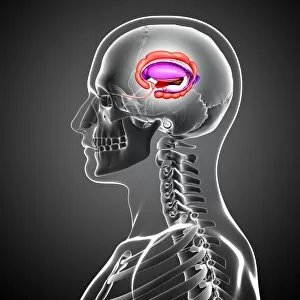
The corpus callosum, a vital structure in the human brain, serves as a bridge connecting the two hemispheres. This intricate network of neural fibers allows for communication and coordination between various regions of our brain. Just like an artist's model, it stands at the center of attention when exploring artworks depicting the limbic system - an area responsible for emotions, memory, and motivation. In mesmerizing brain anatomy artwork, we witness its prominent presence alongside other crucial structures. These captivating illustrations showcase its role in transmitting information across both sides of our brain. As we delve deeper into MRI scans of normal human brains, such as C016/8845 and C016/8850, we can observe this remarkable feature with utmost clarity. With each stroke on canvas or digital rendering capturing its essence flawlessly, these artistic representations emphasize how integral the corpus callosum is to our cognitive functions. It harmoniously connects diverse areas within our brains to ensure seamless integration and efficient processing. Moreover, these artworks also shed light on another significant aspect: the limbic system. By intertwining with this emotional powerhouse within us all, the corpus callosum enables emotions to be shared across both hemispheres. In fascinating depictions like those found in C016/8843 or through visualizations showcasing autonomic nervous systems alongside limbic systems within human bodies – it becomes evident that this connection plays a pivotal role in shaping who we are. Beyond mere illustrations lies advanced technology like DTI MRI scans revealing intricate details about white matter pathways within our brains – including those traversing through the corpus callosum itself. These images provide invaluable insights into how this structure interacts with surrounding regions while highlighting its significance in maintaining overall brain function. Whether portrayed through stunning artwork or captured by cutting-edge medical imaging techniques; one thing remains certain – the corpus callosum holds immense importance in understanding and appreciating the complexity of our incredible minds.