
Cranial Collection
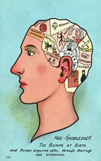
"Cranial Chronicles: Unveiling the Secrets of the Mind's Map" Step into the world exploration, where satire meets phrenology and skull anatomy reigns supreme


All Professionally Made to Order for Quick Shipping
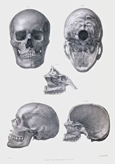
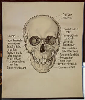
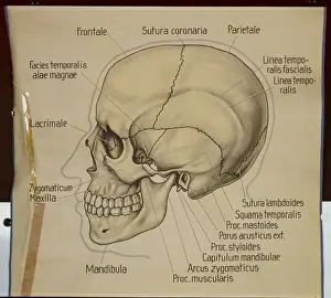
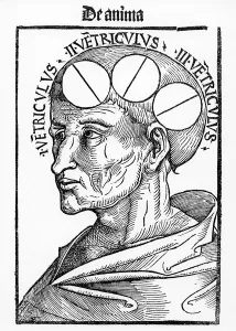
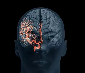
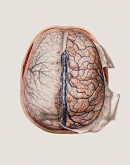
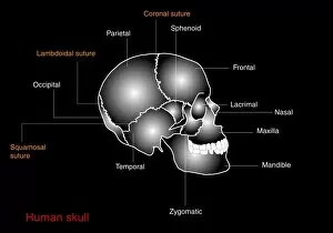
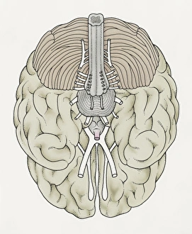
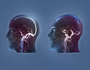
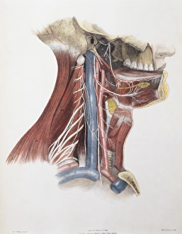
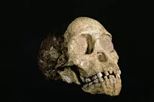
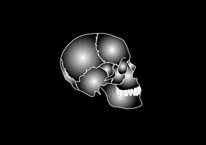
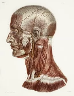
"Cranial Chronicles: Unveiling the Secrets of the Mind's Map" Step into the world exploration, where satire meets phrenology and skull anatomy reigns supreme. Delve into the intricate illustrations of Studio on Saint, as they unravel the mysteries hidden within our craniums. Witness the captivating engravings from "An Operation on the Head, 1577, " a monochromatic masterpiece that takes you back to a time when trepanation was considered cutting-edge medicine. Marvel at its b/w photo counterpart, capturing history frozen in time. Behold lithographic posters showcasing human skulls from different angles – front and side views – reminding us of our own mortality while highlighting their undeniable beauty. These lithos serve as a haunting reminder that beneath our skin lies an intricate network waiting to be explored. Turn your attention to Ms Lat. 7134 fol. 1 Trepanation, extracted from Chirugie de Maitre Rolandi (vellum), offering insight into ancient surgical practices that sought to alleviate cerebral ailments. Let this vellum manuscript transport you through time and space. Explore further with Albertus Magnus' Philosophia Naturalis, revealing cavities within the brain like never before seen. This philosophical journey will challenge your understanding of cognition and leave you pondering over its intricacies long after turning its pages. Marvel at modern medical advancements captured in stunning 3D scans depicting brain aneurysms and cerebral vasculitis - reminders that even today we are still unlocking secrets held within these delicate structures. Finally, gaze upon Opthalmic and maxillary nerves intertwined within the fifth cranial nerve; witness how interconnectedness extends beyond mere physicality into realms yet unexplored by science. Join us on this whimsical journey through cranial wonders - where art meets science, satire dances with phrenology, and knowledge awaits those brave enough to venture inside their own minds.