
Crystallography Collection
Crystallography is a captivating scientific field that unravels the intricate beauty of crystals and their atomic structures


All Professionally Made to Order for Quick Shipping
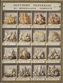
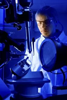
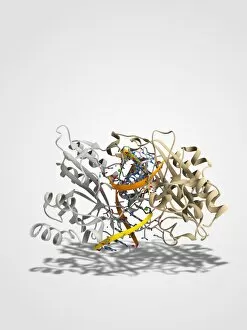
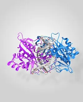
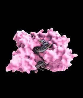
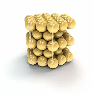
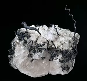
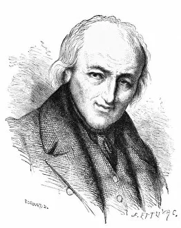
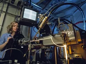
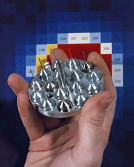
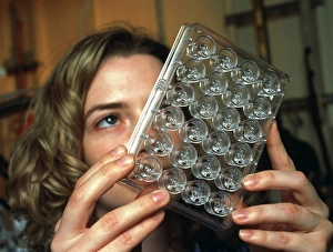
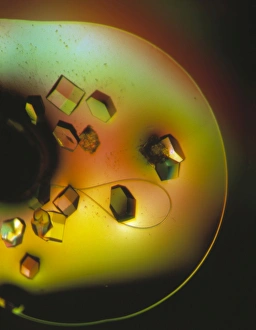
Crystallography is a captivating scientific field that unravels the intricate beauty of crystals and their atomic structures. Dating back to the late 18th century, this engraving titled "Plate 1 from Histoire naturelle? (1789)" serves as a testament to the early exploration of crystallography. Fast forward to modern times, where cutting-edge technologies like the Advanced Light Source synchrotron enable scientists to delve deeper into crystallographic studies. One remarkable discovery in crystallography involves lysozome protein crystals. These microscopic wonders have provided invaluable insights into biological processes and paved the way for breakthroughs in medicine. The mesmerizing image of these crystals showcases their symmetrical patterns and delicate arrangements. Amongst notable figures in this field is William Henry Bragg, an English physicist who founded X-ray crystallography. His pioneering work revolutionized our understanding of crystal structures by utilizing X-rays as a powerful tool for analysis. Bragg's legacy continues to inspire countless researchers worldwide. Intriguingly, even seemingly unrelated inventions find connections with crystallography. Take George Smarts chimney cleansing machine, for instance - its design draws inspiration from the ordered structure found within crystals. This unexpected link highlights how diverse disciplines can intersect and contribute to scientific advancements. The c19th-century depiction aptly captures the essence of historical practices in crystallography when techniques were still evolving but held immense potential for future discoveries. It serves as a reminder of how far we've come since then, thanks to continuous innovation and technological advancements. X-ray crystallography C016 / 3824 represents another milestone achieved through relentless research efforts. By harnessing X-rays' ability to penetrate matter, scientists can obtain detailed images revealing atomic arrangements within various crystalline substances. EcoRV restriction enzyme molecule C014 / 2117 showcases yet another facet of crystallographic investigations - exploring the structures of molecules.