
Cyst Collection
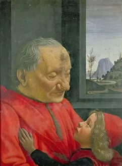
"Cyst: A Tale of Time and Troubles" In the realm of art, an old man and a boy stand frozen in time, their story captured on a 1480s oil painting


All Professionally Made to Order for Quick Shipping
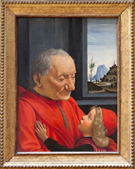
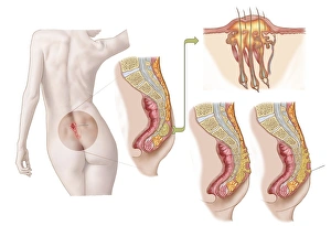
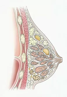
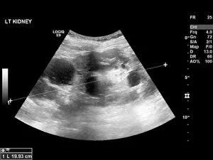
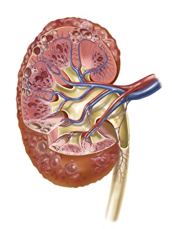
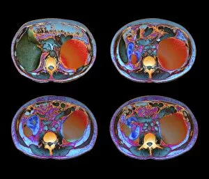
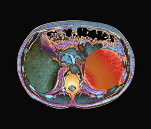
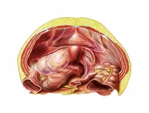
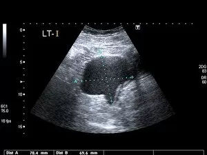
"Cyst: A Tale of Time and Troubles" In the realm of art, an old man and a boy stand frozen in time, their story captured on a 1480s oil painting. Their weathered faces tell tales of wisdom and innocence, as they navigate through life's challenges together. Meanwhile, on another canvas, an illustration reveals the perplexing nature of a dorsal wrist ganglion cyst. Its presence serves as a reminder that even within our bodies, unexpected obstacles can arise. Turning to yet another lithograph, we encounter the face of an ugly man. Though his appearance may be unconventional, it reminds us that beauty lies beyond mere physicality, and is found in resilience and character. As we journey further into history with a c. 1490 oil painting titled "An Old Man and a Boy, " we witness the passage of time etched upon their features. The bond between generations endures despite the trials faced along the way. Venturing into medical illustrations, we come across various manifestations of cysts - from ovarian cysts reaching full development to pilonidal cysts nestled near the natal cleft of buttocks. These images shed light on both common ailments and rare conditions that affect human health. The cross-section illustration showcasing fibroadenoma alongside fibrocystic disease within a human breast emphasizes how intricate our bodies can be. It serves as a reminder to prioritize self-care and seek medical attention when needed. Calcified cysts found within ovaries offer insight into complex reproductive systems while polycystic liver disease presents itself through ultrasound scans - C017 / 8021 revealing its presence in stark detail. Similarly detected by ultrasound scan C017 / 7746 is polycystic kidney disease which highlights how organs can become burdened by multiple fluid-filled sacs over time. Lastly, delving into female anatomy brings us face-to-face with fibroid tumors residing within uteruses - a reminder that even the most intimate parts of our bodies can harbor unexpected challenges.