Fraud Blocker Skip to main content

Cytology Collection (page 32)

Cytology: Exploring the Intricate World of Cells Budding yeast cell: Witness the remarkable process of cellular reproduction as a budding yeast cell emerges

Background imageCytology Collection: Chlamydia, SEM

Chlamydia, SEM
Chlamydia. Coloured scanning electron micrograph (SEM) of chlamydia (Chlamydia sp.) bacteria (green, spherical, centre) in a ruptured cell

Background imageCytology Collection: Glial cell, SEM

Glial cell, SEM
Glial cell. Coloured scanning electron micrograph (SEM) of a cultured glial cell (centre). Glial cells are nervous system cells that provide structural support and protection for neurons (nerve cells)

Background imageCytology Collection: Dead neutrophil white blood cell, SEM

Dead neutrophil white blood cell, SEM
Dead neutrophil white blood cell. Coloured scanning electron micrograph (SEM) of a dead neutrophil (green) surrounded by rod-shaped bacteria (blue). Neutrophils are part of the bodys immune response

Background imageCytology Collection: Human blood cells, SEM

Human blood cells, SEM
Human blood cells, coloured scanning electron micrograph (SEM). Seen here are platelets (small, roundish) and a neutrophil white blood cell (large, whitish)

Background imageCytology Collection: Blood cells in a kidney, SEM

Blood cells in a kidney, SEM
Blood cells in a kidney. Transmission electron micrograph (TEM) of a section through a kidney glomerulus, showing red blood cells (erythrocytes, red) and a white blood cell (leucocyte, blue)

Background imageCytology Collection: Water lily leaf stalk, light micrograph

Water lily leaf stalk, light micrograph
Water lily leaf stalk. Light micrograph of a transverse section through the leaf stalk (petiole) of a water lily (Nymphaea sp.). All aquatic plants (hydrophytes) have a similar structure

Background imageCytology Collection: Pondweed stem, light micrograph

Pondweed stem, light micrograph
Pondweed stem. Light micrograph of a transverse section through the stem of a pondweed (Potamogeton sp.) plant. All aquatic plants (hydrophytes) have a similar stem structure

Background imageCytology Collection: Water milfoil stem, light micrograph

Water milfoil stem, light micrograph
Water milfoil stem. Light micrograph of a transverse section through the stem of the aquatic whorled water milfoil (Myriophyllum verticillatum) plant

Background imageCytology Collection: Water milfoil shoot-tip, light micrograph

Water milfoil shoot-tip, light micrograph
Water milfoil shoot tip. Light micrograph of a transverse section through the shoot-tip of the aquatic whorled water milfoil (Myriophyllum verticillatum) plant

Background imageCytology Collection: Waterweed stem, light micrograph

Waterweed stem, light micrograph
Waterweed stem. Polarised light micrograph of a transverse section through a stem of the aquatic western waterweed (Elodea nuttallii) plant

Background imageCytology Collection: Mistletoe stem, light micrograph

Mistletoe stem, light micrograph
Mistletoe stem. Polarised light micrograph of a transverse section through the stem of a mistletoe (Viscum album) plant. The epidermis (outer layer)

Background imageCytology Collection: Mares tail stem, light micrograph

Mares tail stem, light micrograph
Mares tail stem. Polarised light micrograph of a transverse section through a stem of the aquatic mares tail (Hippuris vulgaris) plant. All aquatic plants (hydrophytes) have a similar stem structure

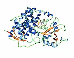
Background imageCytology Collection: Cytochrome P450 molecule

Cytochrome P450 molecule
Cytochrome P450 protein. Molecular model showing the secondary structure of cytochrome P450. This protein plays a crucial role in metabolism in animals (including humans), fungi, plants and bacteria

Background imageCytology Collection: Intestinal smooth muscle cells, TEM

Intestinal smooth muscle cells, TEM
Intestinal smooth muscle cells. Coloured transmission electron micrograph (TEM) of a section through several smooth muscle cells from the intestines

Background imageCytology Collection: Thyroid parafollicular cell, TEM

Thyroid parafollicular cell, TEM
Thyroid parafollicular cell, coloured transmission electron micrograph (TEM). This section has revealed the cells nucleus (light brown)

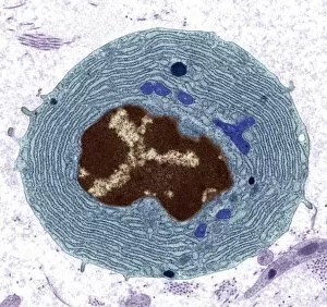
Background imageCytology Collection: Plasma cell, TEM

Plasma cell, TEM
Plasma cell, coloured transmission electron micrograph (TEM). This section has revealed the cells large central nucleus (brown)

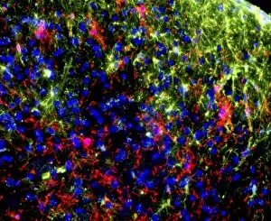
Background imageCytology Collection: Nerve cell trauma response

Nerve cell trauma response. Fluorescent light micrograph of a section through a spinal cord affected by multiple sclerosis (MS)

Background imageCytology Collection: Fibrin strands, artwork

Fibrin strands, artwork
Fibrin strands. Computer artwork depicting a number of fibrin strands with their ends embedded in the membrane of a cell. Fibrin is composed of fibrinogen proteins


All Professionally Made to Order for Quick Shipping

Cytology: Exploring the Intricate World of Cells Budding yeast cell: Witness the remarkable process of cellular reproduction as a budding yeast cell emerges, paving the way for new life. T lymphocytes and cancer cell, SEM C001 / 1679: Delve into the battle between our immune system's T lymphocytes and cancer cells, captured in stunning detail through scanning electron microscopy. Anaesthetic inhibiting an ion channel C015 / 6718: Uncover the secrets behind anesthesia as we observe its impact on ion channels within cells, revealing how it alters our perception of pain. Glial cells, confocal light micrograph: Immerse yourself in the intricate network of glial cells that support and protect neurons in our nervous system, visualized using confocal light microscopy. HeLa cells, light micrograph C017 / 8299: Explore one of the most famous cell lines - HeLa cells - under a microscope to gain insights into their structure and function within biomedical research. Rough endoplasmic reticulum, TEM: Journey deep inside a cell to witness the rough endoplasmic reticulum at work – a vital organelle involved in protein synthesis – revealed through transmission electron microscopy. Glial stem cell culture, light micrograph: Step into the realm of regenerative medicine as we observe glial stem cells cultured in vitro with hopes of unlocking their potential for repairing damaged neural tissue. Dendritic cells, artwork: Marvel at an artistic representation showcasing dendritic cells' crucial role as sentinels of our immune system – capturing antigens and presenting them to other immune cells for recognition and response. Cell structure: Embark on a visual journey exploring various cellular structures that form the building blocks of life itself – from membranes to cytoplasm to nuclei – highlighting their diverse functions.