
Cytoplasmic Extension Collection
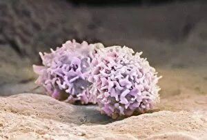
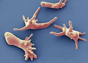
"Cytoplasmic Extension: Unveiling the Dynamic World Within Cells" In this captivating image, an activated macrophage takes center stage


All Professionally Made to Order for Quick Shipping
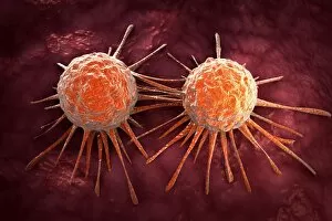
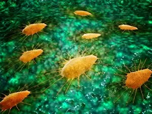
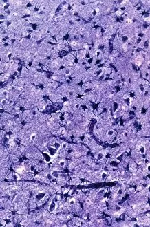
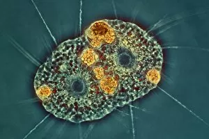
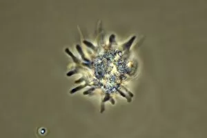
"Cytoplasmic Extension: Unveiling the Dynamic World Within Cells" In this captivating image, an activated macrophage takes center stage, showcasing its remarkable cytoplasmic extensions. These intricate projections emanate from the cell's body, reaching out like tentacles to explore and interact with its surroundings. The scanning electron microscope (SEM) C015/6375 reveals a mesmerizing conceptual image of cancer virus particles lurking in the background. This juxtaposition highlights the crucial role that these cytoplasmic extensions play in immune defense against malignant invaders. Zooming closer into this microscopic realm, we encounter another conceptual image of cancer viruses, emphasizing their potential threat to our cellular harmony. The complexity of their structure serves as a reminder of the challenges faced by our immune system in combating these relentless foes. Shifting gears, we delve into renal corpuscles and their filtration membranes. Here, cytoplasmic extensions take on a different purpose - aiding in kidney function by facilitating efficient filtration processes. This vital interplay between cells and their environment ensures proper waste removal and maintenance of homeostasis within our bodies. Returning to the world of cancer viruses through yet another conceptual image, we witness how they infiltrate healthy cells while evading detection mechanisms, and is at this critical juncture that activated macrophages armed with extensive cytoplasmic extensions come into action – engulfing and neutralizing these harmful intruders before they can wreak havoc on our health. Microscopically observing a group of macrophages further emphasizes their importance as defenders against disease. Their interconnected web-like network showcases how cytoplasmic extensions enable effective communication among immune cells for coordinated responses against pathogens. Meanwhile, glial cells make an appearance under light microscopy – demonstrating yet another facet where cytoplasmic extensions contribute to neural support and protection within our nervous system. Lastly, amoeboid protozoa steal the spotlight under SEM once more, showcasing their own cytoplasmic extensions.