
Daughter Cell Collection
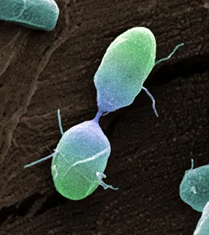
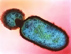
"Daughter Cell: The Miraculous Journey of Life's Replication" Salmonella bacterium dividing


All Professionally Made to Order for Quick Shipping
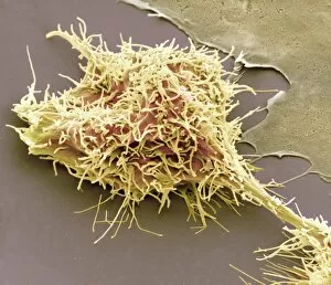
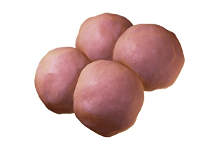
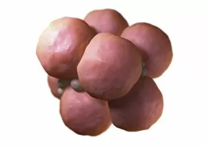
"Daughter Cell: The Miraculous Journey of Life's Replication" Salmonella bacterium dividing, SEM: Witness the intricate process as a Salmonella bacterium splits into two daughter cells, ensuring its survival and propagation. Cell division, SEM: Delve into the microscopic world where cells undergo division, giving rise to identical daughter cells with immense potential for growth and specialization. Dividing brain cancer cells, SEM C014 / 0354: Explore the realm of cellular reproduction within brain cancer cells as they multiply uncontrollably, highlighting the urgency in understanding this complex phenomenon. 4-cell embryo artwork: Marvel at an artist's rendition capturing the delicate stage when a four-cell embryo forms - a crucial step towards embryonic development and future life. Multi-celled embryo artwork: Embark on a visual journey through an artist's interpretation showcasing the awe-inspiring moment when multiple cells unite to form a multi-celled embryo brimming with possibilities. 2-cell embryo artwork: Immerse yourself in an artistic representation that encapsulates the early stages of life as two embryonic cells merge together, setting in motion an incredible chain reaction of growth and differentiation. Cytokinesis artwork: Uncover nature's ingenious mechanism known as cytokinesis through captivating artistry; witness how one cell divides into two separate entities while maintaining their genetic integrity. Cell division artwork: Experience the beauty hidden within cellular replication portrayed by skilled artists who capture this mesmerizing dance between chromosomes during cell division - a symphony of life unfolding before our eyes. Cytokinesis diagram: Grasp the intricacies behind cytokinesis through an illustrative diagram that elucidates how cytoplasmic components are evenly distributed among newly formed daughter cells after nuclear division occurs. Cell division conceptual artwork.