
Daughter Cells Collection
"Daughter Cells: The Miraculous Outcome of Cell Division" Cell division is a fascinating process that allows living organisms to grow, repair, and reproduce


All Professionally Made to Order for Quick Shipping
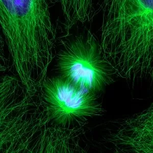
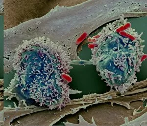
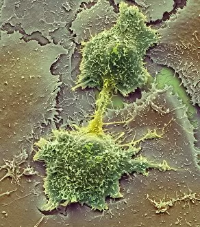
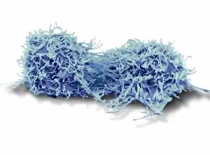
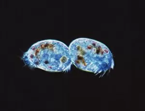
"Daughter Cells: The Miraculous Outcome of Cell Division" Cell division is a fascinating process that allows living organisms to grow, repair, and reproduce. Within the intricate world of microscopic biology, fluorescent micrographs capture the mesmerizing beauty emerging from their parent cell. In one image, we witness a dividing liver cancer cell through scanning electron microscopy (SEM). The detailed view showcases the delicate yet robust nature of these cells as they split into two separate entities. Similarly, another SEM image reveals mouth cancer cells undergoing division - a reminder of the relentless battle against this disease. The transmission electron microscopy (TEM) technique provides us with an even closer look at cellular division. A captivating snapshot displays a dividing cell in all its intricacy and complexity. Meanwhile, another TEM image captures the astonishing sight of dividing cancer cells under SEM C014 / 0362 - highlighting both their destructive potential and resilience. Amongst these images lies an undeniable truth: they can born out of necessity for growth and regeneration. They hold within them immense potential to shape life's course. As we observe yet another SEM image depicting liver cancer cells dividing once more, it serves as a poignant reminder that even amidst chaos and disease, new beginnings can emerge. These captivating glimpses into cellular division remind us that life is constantly evolving on microscopic levels too small for our naked eyes to perceive. Daughter cells symbolize hope; they represent progress in understanding complex diseases like cancer while offering possibilities for innovative treatments. Let us marvel at the wonders hidden within every single divide – where daughter cells come alive – shaping our existence one tiny step at a time.