Fraud Blocker Skip to main content

Deoxyribonucleic Acid Collection

"Unlocking the Blueprint of Life

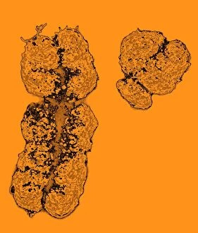
Background imageDeoxyribonucleic Acid Collection: X and Y chromosomes

X and Y chromosomes, coloured scanning electron micrograph (SEM). Humans have 46 chromosomes in total: 23 inherited from the mother and 23 from the father

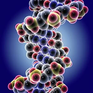
Background imageDeoxyribonucleic Acid Collection: DNA molecule

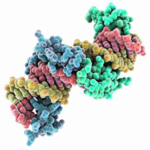
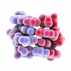
DNA molecule. 3D-computer artwork of a space filling molecular model of deoxyribonucleic acid (DNA)

Background imageDeoxyribonucleic Acid Collection: DNA transcription, molecular model

DNA transcription, molecular model

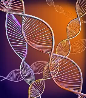
Background imageDeoxyribonucleic Acid Collection: DNA molecule

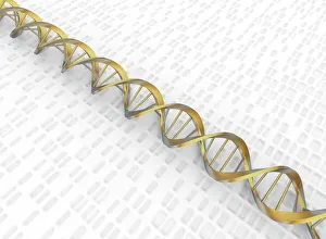
DNA molecule, computer artwork. DNA (deoxyribonucleic acid) is composed of two strands twisted into a double helix

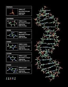
Background imageDeoxyribonucleic Acid Collection: DNA molecule

DNA molecule. Computer artwork of a molecule of DNA (deoxyribonucleic acid) with the chemical formulas of its components

Background imageDeoxyribonucleic Acid Collection: Mitosis, light micrograph

Mitosis, light micrograph
Mitosis. Confocal light micrograph of the stages of mitosis (nuclear division) and cytokinesis (cell division)

Background imageDeoxyribonucleic Acid Collection: Watson and Crick, DNA discovers

Watson and Crick, DNA discovers
Watson and Crick. Caricature of the molecular biologists and discoverers of the structure of DNA James Watson (born 1928, left) and Francis Crick (1916-2004), with their model of a DNA molecule

Background imageDeoxyribonucleic Acid Collection: Herpes virus replicating

Herpes virus replicating

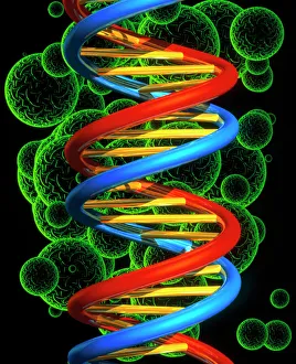
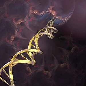
Background imageDeoxyribonucleic Acid Collection: Computer artwork of a beta DNA segment and spheres

Computer artwork of a beta DNA segment and spheres
DNA molecule. Computer artwork of part of a strand of beta DNA (deoxyribonucleic acid) seen on a background of spheres. The spheres may represent bacteria

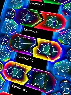
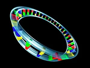
Background imageDeoxyribonucleic Acid Collection: Nucleotide base matrix

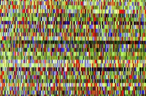
Nucleotide base matrix. Computer artwork depicting a matrix of nucleotide bases: adenine (A), cytosine (C), guanine (G) and thymine (T)

Background imageDeoxyribonucleic Acid Collection: DNA molecule, computer model

DNA molecule, computer model
DNA molecule. Computer artwork of the molecular structure of DNA (deoxyribonucleic acid). The DNA molecule is composed of two strands twisted into a double helix

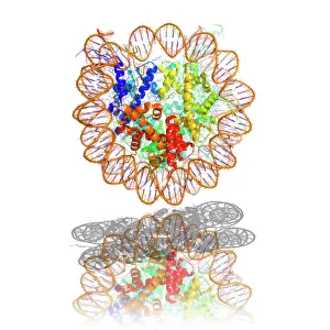
Background imageDeoxyribonucleic Acid Collection: Nucleosome molecule

Nucleosome molecule, computer model. A nucleosome is a subunit of chromatin, the substance that forms chromosomes

Background imageDeoxyribonucleic Acid Collection: DNA molecule, abstract image

DNA molecule, abstract image
DNA molecule. Abstract computer artwork of a view along the inside of a molecule of DNA (deoxyribonucleic acid). DNA contains sections called genes that encode the bodys genetic information

Background imageDeoxyribonucleic Acid Collection: DNA nucleosome, molecular model

DNA nucleosome, molecular model
DNA nucleosome. Molecular model of a nucleosome, the fundamental repeating unit used to package DNA (deoxyribonucleic acid) inside cell nuclei

Background imageDeoxyribonucleic Acid Collection: DNA molecule, artwork

DNA molecule, artwork
DNA molecule. Computer artwork of a double stranded DNA (deoxyribonucleic acid) molecule amongst clouds of swirling gas. DNA is composed of two strands twisted into a double helix

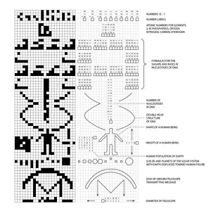
Background imageDeoxyribonucleic Acid Collection: Arecibo message and decoded key C016 / 6817

Arecibo message and decoded key C016 / 6817
Arecibo message and decoded key. Diagram showing the binary transmission (left) known as the Arecibo message, with the message decoded and labelled at centre and right

Background imageDeoxyribonucleic Acid Collection: Chloroplast structure, artwork

Chloroplast structure, artwork
Chloroplast structure. Artwork showing the internal structure of chloroplasts, the organelles in plant cells responsible for photosynthesis

Background imageDeoxyribonucleic Acid Collection: DNA Double Helix with Autoradiograph

DNA Double Helix with Autoradiograph
Conceptual computer illustration of the DNA double helix together with a graphic representation of an autoradiograph display

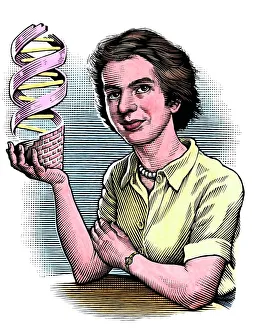
Background imageDeoxyribonucleic Acid Collection: Rosalind Franklin, British chemist

Rosalind Franklin, British chemist
Rosalind Franklin (1920-1958), British chemist and X-ray crystallographer, holding a model of DNA (deoxyribonucleic acid)

Background imageDeoxyribonucleic Acid Collection: Zinc fingers bound to a DNA strand

Zinc fingers bound to a DNA strand, molecular model. The double helix of DNA (deoxyribonucleic acid, red and yellow) is seen here with two Zif268 proteins (blue and green)

Background imageDeoxyribonucleic Acid Collection: HeLa cells, light micrograph C013 / 4774

HeLa cells, light micrograph C013 / 4774
HeLa cells. Multi-photon fluorescence light micrograph of a group of cultured HeLa cells, showing the cell nuclei, which contain the cells genetic information (DNA, red)

Background imageDeoxyribonucleic Acid Collection: Chromosome

Chromosome. Computer artwork of a condensed chromosome

Background imageDeoxyribonucleic Acid Collection: Dividing cells

Dividing cells. Computer artwork of a spiral of cells during mitosis (nuclear division). During mitosis two daughter nuclei are formed from one parent nucleus

Background imageDeoxyribonucleic Acid Collection: Anti-cancer drug binding to DNA, AFM

Anti-cancer drug binding to DNA, AFM
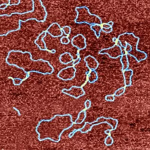
Drug-DNA complexes. Coloured atomic force micrograph (AFM) of plasmids (blue) of DNA (deoxyribonucleic acid) bound to the anti-cancer drug ditercalinium

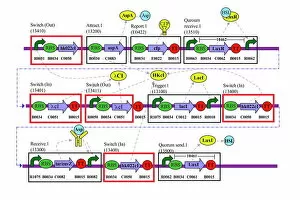
Background imageDeoxyribonucleic Acid Collection: Genetic circuit diagram

Genetic circuit diagram. Genetic circuits are used by synthetic biologists to design genetically modified bacteria cells

Background imageDeoxyribonucleic Acid Collection: DNA autoradiogram, artwork

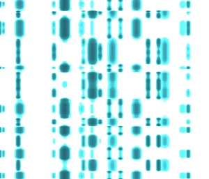
DNA autoradiogram, artwork. Autoradiograms show the order of nucleotide bases (basic building blocks) in a sample of DNA (deoxyribonucleic acid)

Background imageDeoxyribonucleic Acid Collection: Computer artwork of DNA replication

Computer artwork of DNA replication
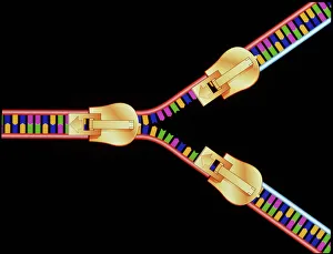
DNA replication. Computer artwork depicting DNA (deoxyribonucleic acid) replication. This segment of DNA is being " unzipped" to form a Y-shaped replication fork

Background imageDeoxyribonucleic Acid Collection: Grapevine genome sequencing

Grapevine genome sequencing. Data from a gelelectrophoresis experiment to sequence the PinotNoir grape ( Vitis sp. ) genome

Background imageDeoxyribonucleic Acid Collection: Full set of male chromosomes, SEM

Full set of male chromosomes, SEM
Set of human chromosomes, coloured scanning election micrograph (SEM). Chromosomes are a packaged form of the genetic material deoxyribonucleic acid (DNA)

Background imageDeoxyribonucleic Acid Collection: Z-DNA tetramer molecule C015 / 6557

Z-DNA tetramer molecule C015 / 6557
Z-DNA (deoxyribonucleic acid) tetramer, molecular model. DNA is composed of two strands twisted into a double helix. This is a tetramer of the molecule, containing four strands

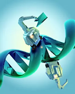
Background imageDeoxyribonucleic Acid Collection: DNA repair using nanobots

DNA repair using nanobots. Computer artwork depicting the possibility of using robots at an atomic scale (nanobots) to repair damaged DNA (deoxyribonucleic acid), the bodys genetic code

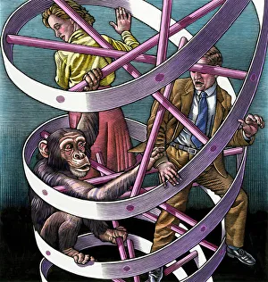
Background imageDeoxyribonucleic Acid Collection: Shared DNA in humans and chimps, art

Shared DNA in humans and chimps, art
Shared DNA between humans and chimps, conceptual artwork. The humans look surprised to see the chimp so close to them in the double helix of DNA (deoxyribonucleic acid)

Background imageDeoxyribonucleic Acid Collection: Mitochondrial DNA

Mitochondrial DNA. Computer artwork of the genetic material (DNA, deoxyribonucleic acid) found in the cell structures called mitochondria

Background imageDeoxyribonucleic Acid Collection: DNA analysis

DNA analysis, negative image. Graphs showing the results of DNA (deoxyribonucleic acid) sequencing

Background imageDeoxyribonucleic Acid Collection: X and Y chromosomes

X and Y chromosomes, computer artwork. Humans have 46 chromosomes in total: 23 inherited from the mother and 23 from the father. These sex chromosomes determine the individuals gender

Background imageDeoxyribonucleic Acid Collection: AI IMAGE - Portrait of Rosalind Franklin, 1940s, (2023). Creator: Heritage Images

AI IMAGE - Portrait of Rosalind Franklin, 1940s, (2023). Creator: Heritage Images
AI IMAGE - Portrait of Rosalind Franklin, 1940s, (2023)

Background imageDeoxyribonucleic Acid Collection: AI IMAGE - Portrait of Rosalind Franklin, 1940s, (2023). Creator: Heritage Images

AI IMAGE - Portrait of Rosalind Franklin, 1940s, (2023). Creator: Heritage Images
AI IMAGE - Portrait of Rosalind Franklin, 1940s, (2023)

Background imageDeoxyribonucleic Acid Collection: TFAM transcription factor bound to DNA C015 / 7059

TFAM transcription factor bound to DNA C015 / 7059
TFAM transcription factor bound to DNA, molecular model. Human mitochondrial transcription factor A (TFAM, green) bound to a strand of DNA (deoxyribonucleic acid, blue and pink)

Background imageDeoxyribonucleic Acid Collection: DNA by tunnelling microscope

DNA by tunnelling microscope
False-colour scanning tunnelling micrograph (STM) of DNA. A sample of uncoated, double-stranded DNA was dissolved in a salt solution & deposited on graphite prior to being imaged in air by the STM

Background imageDeoxyribonucleic Acid Collection: DNA strands, illustration

DNA strands, illustration
DNA strands. Computer illustration showing the structure of double stranded DNA (deoxyribonucleic acid) molecules. DNA is composed of two strands twisted into a double helix

Background imageDeoxyribonucleic Acid Collection: Microscopic view of DNA

Microscopic view of DNA

Background imageDeoxyribonucleic Acid Collection: DNA structure, artwork C017 / 7218

DNA structure, artwork C017 / 7218
DNA structure. Computer artwork showing the structure of a double stranded DNA (deoxyribonucleic acid) molecule (right) and its components (left)

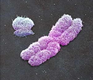
Background imageDeoxyribonucleic Acid Collection: Male sex chromosomes, SEM

Male sex chromosomes, SEM
Male sex chromosomes. Coloured scanning electron micrograph (SEM) of human X (centre) and Y (upper left) sex chromosomes. Each chromosome has replic- ated to form two identical strands (chromatids)

Background imageDeoxyribonucleic Acid Collection: High-contrast direct DNA image, TEM

High-contrast direct DNA image, TEM
High-contrast direct DNA image. Coloured transmission electron micrograph (TEM) of the first high-contrast direct image of a bundle (fibre) of strands of DNA (deoxyribonucleic acid)

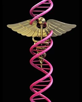
Background imageDeoxyribonucleic Acid Collection: Caduceus with DNA, artwork C013 / 9990

Caduceus with DNA, artwork C013 / 9990
Caduceus with DNA. Computer artwork of the Caduceus symbol entwined by a strand of DNA (deoxyribonucleic acid)

Background imageDeoxyribonucleic Acid Collection: Male Human Sex Chromosomes X and Y SEM C013 / 5127

Male Human Sex Chromosomes X and Y SEM C013 / 5127
Male Human Sex Chromosomes X and Y (Pair 23), scanning electron micrograph (SEM). There are 23 pairs of chromosomes in most normal human cells

Background imageDeoxyribonucleic Acid Collection: Creation of oil using designer microbes

Creation of oil using designer microbes. Conceptual computer artwork depicting an oil well pump in a petri dish, representing the use of artificially created micro-organisms to produce oil

Background imageDeoxyribonucleic Acid Collection: Creation of artificial life, artwork

Creation of artificial life, artwork
Creation of artificial life. Conceptual computer artwork depicting the creation of a new life-form in a petri dish

Background imageDeoxyribonucleic Acid Collection: DNA autoradiogram and face

DNA autoradiogram and face
MODEL RELEASED. DNA autoradiogram. Computer artwork of a DNA (deoxyribonucleic acid) autoradiogram superimposed over a womans face

Background imageDeoxyribonucleic Acid Collection: DNA electrophoresis gels, artwork

DNA electrophoresis gels, artwork
DNA electrophoresis. Computer artwork of agarose electrophoresis gels. Each gel reveals different fragments of DNA. The fragments are separated by applying an electric current across the gel

Background imageDeoxyribonucleic Acid Collection: Genetic security

Genetic security. Conceptual computer artwork of a strand of DNA (deoxyribonucleic acid) locked inside a padlock. This may represent the protection of an individuals genetic code from exploitation

Background imageDeoxyribonucleic Acid Collection: DNA

DNA, computer artwork. DNA (deoxyribonucleic acid) consists of two strands (yellow) of sugar phosphates forming a double helix

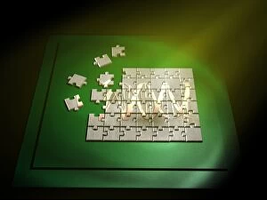
Background imageDeoxyribonucleic Acid Collection: Nude jigsaw

Nude jigsaw
DNA jigsaw. Conceptual computer artwork of a DNA (deoxyribonucleic acid) molecule formed by pieces of a jigsaw puzzle

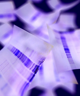
Background imageDeoxyribonucleic Acid Collection: Genetic sequence

Genetic sequence. Printout of the genetic code of a single strand of DNA (deoxyribonucleic acid)

Background imageDeoxyribonucleic Acid Collection: Bacterial cell structure, artwork

Bacterial cell structure, artwork
Bacterial cell structure. Computer artwork showing the cell structure and components (organelles) of a typical rod-shaped bacteria (bacillus). Not all bacteria have a flagellum (long, tail-like)

Background imageDeoxyribonucleic Acid Collection: Smallpox virus particle, TEM

Smallpox virus particle, TEM
Smallpox virus particle. Coloured transmission electron micrograph (TEM) of a Variola major virus particle

Background imageDeoxyribonucleic Acid Collection: Francis Harry Compton Crick, British microbiologist, c1962

Francis Harry Compton Crick, British microbiologist, c1962. Francis Crick (1916-2004) discovered the molecular structure of DNA

Background imageDeoxyribonucleic Acid Collection: Illustration of structure of human Deoxyribonucleic acid (DNA)

Illustration of structure of human Deoxyribonucleic acid (DNA)

Background imageDeoxyribonucleic Acid Collection: Conceptual image of a telomere

Conceptual image of a telomere. A telomere is a region of the DNA sequence at the end of a chromosome. Their function is to protect the ends of the chromosome from degradating

Background imageDeoxyribonucleic Acid Collection: Cell nucleus with chromosome

Cell nucleus with chromosome. The cell nucleus helps control eating, movement, and reproduction

Background imageDeoxyribonucleic Acid Collection: Microscopic view of DNA binding

Microscopic view of DNA binding

Background imageDeoxyribonucleic Acid Collection: Microscopic view of telomeres highlighted at the tips of chromosome

Microscopic view of telomeres highlighted at the tips of chromosome. A telomere is a region of the DNA sequence at the end of a chromosome

Background imageDeoxyribonucleic Acid Collection: Multiphoton fluorescence image of HeLa cells

Multiphoton fluorescence image of HeLa cells with cytoskeletal microtubules (magenta) and DNA (cyan)

Background imageDeoxyribonucleic Acid Collection: Conceptual image of chromosomes inside the blood stream

Conceptual image of chromosomes inside the blood stream

Background imageDeoxyribonucleic Acid Collection: Conceptual image of a telomere showing DNA structure

Conceptual image of a telomere showing DNA structure. A telomere is a region of the DNA sequence at the end of a chromosome. Their function is to protect the ends of the chromosome from degradation

Background imageDeoxyribonucleic Acid Collection: Microscopic view of chromosome

Microscopic view of chromosome

Background imageDeoxyribonucleic Acid Collection: Stylized view of strands of human DNA

Stylized view of strands of human DNA or deoxyribonucleic acid

Background imageDeoxyribonucleic Acid Collection: Microscopic view of cancer cells

Microscopic view of cancer cells. Cancer occurs when a cells gene mutations make the cell unable to correct DNA damage

Background imageDeoxyribonucleic Acid Collection: Microscopic view of pancreatic cancer cell

Microscopic view of pancreatic cancer cell

Background imageDeoxyribonucleic Acid Collection: Microscipic view of pancreatic cancer cells

Microscipic view of pancreatic cancer cells

Background imageDeoxyribonucleic Acid Collection: Conceptual image of chromosome

Conceptual image of chromosome

Background imageDeoxyribonucleic Acid Collection: Conceptual image of DNA

Conceptual image of DNA

Background imageDeoxyribonucleic Acid Collection: Cluster of DNA strands

Cluster of DNA strands of human DNA or deoxyribonucleic acid

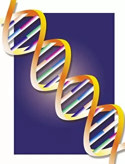
Background imageDeoxyribonucleic Acid Collection: Artwork of DNA structure

Artwork of DNA structure

Background imageDeoxyribonucleic Acid Collection: Stochastic gene expression, illustration C018 / 0906

Stochastic gene expression, illustration C018 / 0906
Stochastic gene expression, illustration. Every cell in an organism contains every single gene that makes up the organisms genome. However, they are not all active (expressed) in each cell

Background imageDeoxyribonucleic Acid Collection: DNA transcription, illustration C018 / 0900

DNA transcription, illustration C018 / 0900
DNA (deoxyribonucleic acid) transcription

Background imageDeoxyribonucleic Acid Collection: Tumour suppressor protein and DNA C017 / 3647

Tumour suppressor protein and DNA C017 / 3647
Tumour suppressor protein and DNA. Computer artwork showing a molecule of the tumour suppressor protein p53 (blue and pink) bound to a molecule of DNA (deoxyribonucleic acid, yellow and orange)

Background imageDeoxyribonucleic Acid Collection: TATA box-binding protein complex C017 / 7082

TATA box-binding protein complex C017 / 7082
TATA box-binding protein complex. Molecular model showing a TATA box-binding protein (TBP, green) complexed with a strand of DNA (deoxyribonucleic acid, yellow) and transcription factor IIB

Background imageDeoxyribonucleic Acid Collection: DNA molecule, artwork C017 / 7217

DNA molecule, artwork C017 / 7217
DNA molecule. Computer artwork showing a double stranded DNA (deoxyribonucleic acid) molecule. DNA is composed of two strands twisted into a double helix

Background imageDeoxyribonucleic Acid Collection: DNA molecule, artwork C017 / 0616

DNA molecule, artwork C017 / 0616
DNA molecule. Computer artwork looking along the interior of a double stranded DNA (deoxyribonucleic acid) molecule. DNA is composed of two strands twisted into a double helix

Background imageDeoxyribonucleic Acid Collection: Telemedicine, conceptual image C017 / 7590

Telemedicine, conceptual image C017 / 7590
Telemedicine, conceptual image

Background imageDeoxyribonucleic Acid Collection: TATA box-binding protein complex C017 / 7088

TATA box-binding protein complex C017 / 7088
TATA box-binding protein complex. Molecular model showing a TATA box-binding protein (TBP, green) complexed with a strand of DNA (deoxyribonucleic acid, yellow) and transcription factor IIB

Background imageDeoxyribonucleic Acid Collection: Genetics research, conceptual artwork C017 / 7410

Genetics research, conceptual artwork C017 / 7410
Genetics research. conceptual computer artwork

Background imageDeoxyribonucleic Acid Collection: DNA molecule, artwork C017 / 0615

DNA molecule, artwork C017 / 0615
DNA molecule. Computer artwork looking along the interior of a double stranded DNA (deoxyribonucleic acid) molecule. DNA is composed of two strands twisted into a double helix


All products are expertly crafted, using premium materials, tailored to your specifications and promptly shipped

"Unlocking the Blueprint of Life: Exploring the Marvels of Deoxyribonucleic Acid" From its iconic double helix structure to its role as the carrier of genetic information, the DNA molecule stands at the core of life's intricate tapestry. The X and Y chromosomes, known for determining our biological sex, are just a fraction of what this remarkable molecule encompasses. DNA transcription, a process where genetic instructions are converted into RNA molecules, allows for protein synthesis and ultimately shapes our traits. This molecular model unveils the intricacies involved in this vital mechanism. Watson and Crick's groundbreaking discovery in 1953 revolutionized biology forever. Their unraveling of DNA's structure paved the way for countless scientific advancements that continue to shape our understanding today. Mitosis, captured through a light micrograph, showcases how DNA faithfully replicates itself during cell division. This fundamental process ensures growth and repair within living organisms. In this computer artwork featuring a beta DNA segment and spheres representing nucleotides, we witness science merging with artistry to depict both complexity and beauty entwined within every strand. The nucleotide base matrix acts as an essential foundation upon which our genetic code is built. Its precise arrangement determines everything from eye color to disease susceptibility—a testament to nature's meticulous design. A computer model showcasing the three-dimensional structure of a DNA molecule further emphasizes its elegance. Every twist and turn holds invaluable information that defines who we are at our very core. Nucleosomes—molecular complexes formed by wrapping DNA around proteins—play a crucial role in organizing chromatin structures within cells. This detailed molecular model highlights their significance in gene regulation and genome stability. An abstract image captures the essence of a DNA molecule—an enigmatic dance between orderliness and randomness that underpins all life forms on Earth. It symbolizes both unity among species yet individuality within each organism. As we delve deeper into understanding deoxyribonucleic acid, we unravel the secrets of our existence.

© Copyright Media Storehouse, All Rights Reserved 2003 - 2024
Facebook     Pinterest     100% Payment Secure     Reviews IO